Biomedical Engineering Reference
In-Depth Information
optical microscopy (OM) [Plieva et al., 2006b; Vlierberghe et al., 2007], scanning
electron microscopy (SEM) [Dubruel et al., 2007; Plieva et al., 2004, 2006a; Vlier-
berghe et al., 2007], environmental scanning electron microscopy (ESEM) [Plieva
et al., 2005, 2006b], confocal microscopy (CM) [Dubruel et al., 2007; Plieva et al.,
2007a] and microcomputed tomography [Dubruel et al., 2007; Vlierberghe et al.,
2007 ].
The cryogel backbone was colored with a dye or fl uorescein - probe to be
visible in OM and CM, respectively. The dye, Cibacrone Blue, was bound to the
network of PVA to make the structure of PVA-MG visible in OM studies showing
large interconnected pores surrounded by dense and microporous pore walls
(Figure 14.13a). Gelatin cryogel scaffolds studied by optical microscopy showed
the absence of “skin layer” which could be a problem for cell in-growth studies
(a)
(b)
(c)
(d)
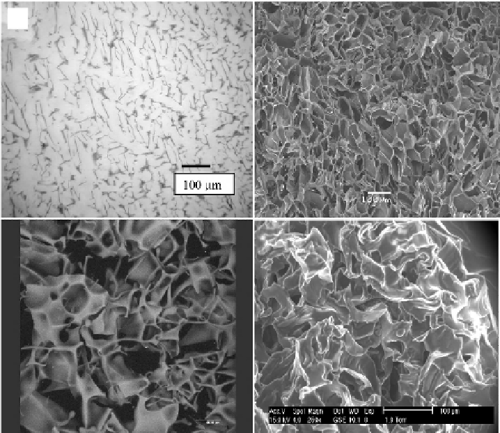
Figure 14.13.
Porous structure of MGs visualized by different techniques: optical microscopy,
OM (a), confocal microscopy, CM (b), scanning electron microscopy, SEM (c) and environmen-
tal scanning electron microscopy, ESEM (d). OM (a) is presented for PVA-MGs and reproduced
from [Plieva et al., 2006b] with permission. CM (b) presented for pAAm-MGs and reproduced
from [Plieva et al., 2006c] with permission. SEM (c) is presented for dextran-MA-MGs and re-
produced from [Plieva et al., 2007a] with permission. The ESEM (d) is presented for PVA-MGs
and reproduced from [Plieva et al., 2006b] with permission.


Search WWH ::

Custom Search