Biomedical Engineering Reference
In-Depth Information
PO
3-
(b)
A
2-
(s)
CO
3
OH
-
(s)
PO
3-
(s)
B
CO
2-
(b)
C
D
E
4000
3500
1600 1400
Wavenumber (cm
-1
)
1250
1000
750
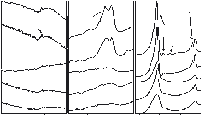
Figure 11.19.
FTIR spectra divided in the main areas of interest of the following samples: A)
HA (0% Si), B) HASi25 (2,5% Si), C) HASi50 (5% Si), D) HASi75 (7,5% Si) and E) HASi100 (10%
Si). The target ablation was carried out with an ArF excimer laser (193 nm), operating at 175 mJ
and a pulse repetition rate of 10 Hz. The fi lms were deposited in a water vapour atmosphere
(0,45 mbar), and the Si and Ti substrates were maintained at 460 °C during the fi lm growth.
From [Solla, 2007].
3. a peak at 940 cm
− 1
present in all the samples except the Si free one (HA),
iv) a small shoulder at 690 cm
− 1
and,
4. a small peak at 670 cm
− 1
for samples with high Si content. These phenom-
ena have been reported before by many authors [Gibson, 1999; Botelho,
2002 ; Arcos, 2004 ].
Figure 11.20 shows the XRD spectra of the HA, HASi50, and HASi100 coatings.
All of the coatings produced exhibit a crystalline structure and present exactly
the same refl ection peaks as the reference coating of pure HA, thus it is clear that
all the coatings have a hydroxyapatite structure and no secondary phases were
formed due to the Si substitution. The only difference that arises is that, when the
Si content is increased, XRD refl ections become less intense. This phenomenon
indicates a progressive loss of cristallinity along with the Si substitution, a fact
that is coherent with the FTIR analysis aforementioned.
The chemical surface characterization of the Si-HA coatings was performed
by XPS analysis which provides very useful information on the Si bonding envi-
ronment. The quantifi cation of the Si transferred to the coating from the initial
ablation target is summarised in Table 11.3. It is clear that as the concentration of
Si in the ablation target increases, the quantity of Si transferred to the coating

Search WWH ::

Custom Search