Biomedical Engineering Reference
In-Depth Information
traditional PLGA) and ease of degradability, it has been considered as a new
generation of tissue engineering scaffold for numerous tissue engineering com-
posite applications especially for bone and articular cartilage.
7.3.3.2 Nanostructured and Injectable Hydrogels as Bone Tissue Engi-
neering Scaffolds.
Polymeric hydrogels have excellent biocompatibility which
makes them useful in orthopedic applications (such as for injectable bone repair
and cartilage reconstruction) [100]. Natural hydrogels (such as collagen and gela-
tin) are the main components of extra cellular matrices (ECM) of mammalian
tissues including bone, cartilage, and so on [101]. Through a technique called elec-
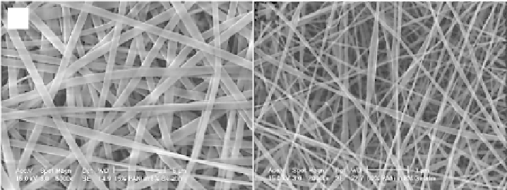
trospinning, nanofi brous scaffolds [104] can be successfully created from various
synthetic and natural polymers such as gelatin and collagen (Figure 7.9).
Several studies have shown that protein and cell interactions are promoted
by nanofi brous scaffolds that biomimic the ECM [102-103]. On the other hand,
studies have demonstrated that using self-assembling peptide KLD-12 hydrogels
for encapsulating chondrocytes can support chondrocyte differentiation and
promote the synthesis of a cartilage-like extracellular matrix for cartilage repair
[105] .
In addition, various synthetic hydrogels such as poly(2-hydroxyethyl methac-
rylate) (pHEMA) were modifi ed to have nanofeatures for orthopedic applica-
tions. For example, some studies have created an injectable nanostructured
pHEMA-hydrogel scaffold which incorporates novel self-assembled helical
rosette nanotubes (HRNs) into hydrogels to fi ll bone fractures and repair bone
defects [106]. The nanoscale, tubular architecture of HRNs on and in hydrogels
can provide a topography that improves protein adsorption and an environment
(a)
(b)
Figure 7.9.
Scanning electron microscopy images of electrospun polyaniline-contained
gelatin nanofi bers. (A) polyaniline-gelatin blend fi bers with ratios of 15 : 85 and (B)
polyaniline-gelatin blend fi bers with ratios of 60 : 40. This Fig. shows that the electrospun fi bers
are homogeneous while 60 : 40 fi bers were electrospun with beads [104]. Scale bars = 5
μ
m for
(A) and 1
μ
m for (B).
Search WWH ::

Custom Search