Biomedical Engineering Reference
In-Depth Information
Uncoated tantalum
Conventional HA coated tantalum
Nanocrystalline HA coated tantalum Nanocrystalline HA coated tantalum
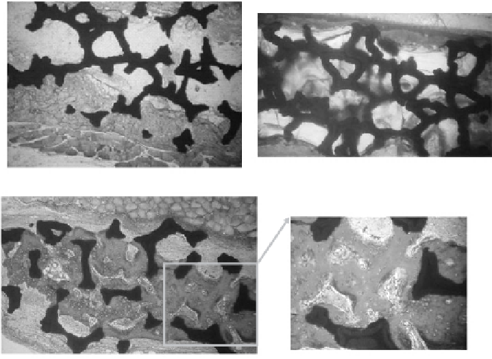
Figure 7.4.
Histology of rat calvaria after 6 weeks of implantation of uncoated tantalum,
conventional HA coated tantalum and nanocrystalline HA coated tantalum. Greater amounts
of new bone formation occurred in the rat calvaria with implanting nanocrystalline HA coated
tantalum compared to uncoated and conventional HA coated tantalum. Red represents new
mineralized bone and blue represents unmineralized tissue. Adapted and redrawn from [131].
(See color insert.)
or cavities since it showed good tissue incorporation, high biocompatibility and
rapid osseointegration [56].
To demonstrate the versatility of nano ceramics, similar tendencies also
appeared for nanophase alumina, zirconia and titania. A 51% increase in osteo-
blast adhesion and a 235% decrease in fi broblast adhesion in a four-hour period
were observed on alumina as grain size decreased from 167 to 24 nm [16]. More-
over, osteoblast adhesion increased by 146% and 200% on nanophase zirconia
(23 nm) and titania (32 nm) compared to microphase zirconia (4.9
μ
m) and titania
(4.1
m), respectively, when normalized to projected surface area [57]. Further-
more, increased collagen synthesis, alkaline phosphatase activity and calcium
mineral deposition by osteoblasts were observed on nanophase zirconia, titania
and alumina (theta + delta crystalline phase) compared to conventional equiva-
lents [48,57] .
To determine the role of nano ceramics in fi ghting implant infection, vari-
ous bacteria functions were recently investigated on nanophase ceramics. For
instance,
Staphylococcus epidermidis
(a common bacterium in human skin)
μ

Search WWH ::

Custom Search