Biomedical Engineering Reference
In-Depth Information
0.5
mm
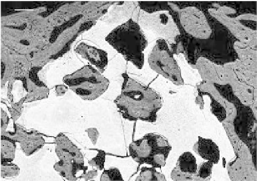
Figure 4.13.
SEM observations showing the newly-formed bone in close contact with MBCP
macropores.
lower in tibial sites than in the ilium or humeral sites in dogs (p
0.05), whereas
no difference was observed between any sites in rabbits. No difference was found
between the ilium or humerus with respect to global haematopoietic cell abun-
dance and count for every lineage. The overall quantity of haematopoietic cells
was signifi cantly lower in dogs than in rabbits for all sites (p
<
0.05).
Three-dimensional imaging made possible an overall examination of the im-
plants and showed a bony formation bridging the whole length of the defect in all
implants. 2D imaging showed that newly-formed bone repartition was homoge-
neous in three implants and relatively heterogeneous in others. In the latter, new
bone was observed essentially around the implant at the initial collagen mem-
brane location. The new bone formation was observed both around the MBCP®
granules and in the macropores with direct contact (Figure 4.13).
With both polarised light microscopy and SEM using BSE, the bone
appeared well-mineralised and organised. Lamellar bone was observed directly
in contact with the MBCP granules without fi brous interposition. From each
host bone, the new bone formation had developed bone extremities from the
defect to the core with osseous continuity. However, the bone ingrowth was
not homogeneous according to the repartition in the axial section. In three im-
plants, new bone was formed both in the centre and the periphery of the implant,
and the MBCP granules appeared totally integrated. In the other three, newly-
formed bone was observed mainly on the periphery of the osseous defects. The
border of the defect was fi lled with compact bone with dense, lamellar structures
with few spaces for vessels and soft tissue. The center of the defect for these three
implants was fi lled with few osseous trabeculae, but large amounts of haemato-
poietic precursors and blood cells were observed. Some granules had no close
contact with new bone formation but were associated with the considerable cel-
lular content of the haematopoietic cells. This trend for peripheral trabecular
<
Search WWH ::

Custom Search