Biomedical Engineering Reference
In-Depth Information
EPI-Mode
Single pass
Wavefront in the
pupil plane
Specimen
Point
source
AFP
NFP
FIGuRE 5.1
Let
:.Specimen-induced.aberrations.and.their.efect.in.epi-coniguration..Because.of.the.variation.
in.refractive.index.within.the.specimen,.the.actual.focal.position.(AFP).of.the.focal.spot.can.be.diferent.from.the.
nominal. focal. position. (NFP)..
Right
:. Wavefront. aberrations. can. be. measured. in. the. pupil. plane. of. the. lens. in. a.
single.pass.coniguration.when.a.point.source.is.placed.in.the.nominal.focal.spot.position..he.measured.wavefront.
information.allows.one.to.model.the.position.and.shape.of.the.focal.spot.in.the.equivalent.epi-coniguration.
Bottom
surface of
cover slip
76
µ
m
Should be flat!
6
µ
m
Top surface
of slide
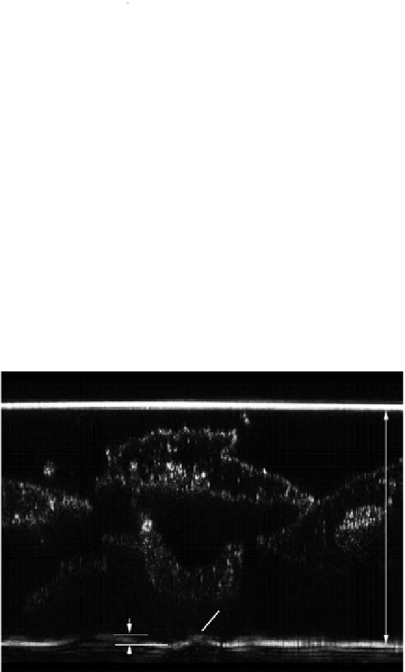
FIGuRE 5.2
Illustration. of. the. specimen-induced. distortions:. confocal. axial. X/Z. scan. of. cheek. cells. above. the.
interface.between.embedding.medium.and.the.microscope.slice..he.relection.from.the.interface.is.marked..In.the.
scan,.because.of.the.discussed.efect,.the.interface.appears.to.be.bent.even.though.the.object.is.known.to.be.lat..From.
Pawley,.J..(2002)..Limitations.on.optical.sectioning.in.live-cell.confocal.microscopy..
Scanning 24
,.241-246,.©.FAMS.Inc.
the.intensity.sampled.at.the.AFP.into.a.3D.data.set.using.the.information.that.was.actually.collected.at.
the.NFP..his.leads.to.local.spatial.distortions.in.the.data.set.and.can.cause.measurement.inaccuracies..
A.striking.example.was.presented.by.Pawley.(2002)..An.image.from.this.article.is.shown.in.Figure.5.2,.
where.the.relection.image.from.the.lat.interface.between.the.embedding.medium.of.the.specimen.and.
the.microscope.slide.is.distorted.because.of.the.refractive.index.structure.of.the.cell.above.