Biomedical Engineering Reference
In-Depth Information
PSF
PSF
(a)
(b)
FIGuRE 17.26
he.images.and.PSF.without.(a).and.with.(b).correction.for.a.cycle.14.fruit.ly.embryo.with.green.
luorescent.protein-polo.at.the.depth.of.83.μm..he.scale.bar.is.2.μm.
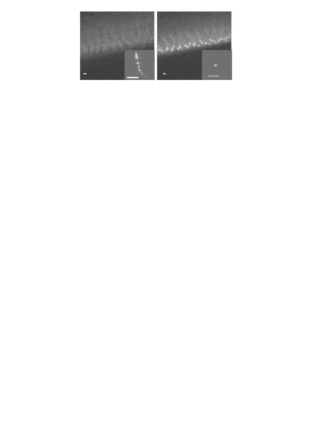
are.captured.at.the.depth.of.83.μm.as.shown.in.Figure.17.26..he.GFP-polo-labeled.centrosomes.can.be.
observed.clearly.ater.correction.but.cannot.be.observed.before.correction..he.size.of.the.PSF.decreases.
from.1.7.to.0.21.μm..he.Strehl.ratio.calculated.based.on.the.PSF.shows.an.increase.from.3.3.×.10
−3
.to.0.7..
he.penetration.depth.for.live.imaging.of.a.fruit-ly.embryo.is.tested.by.performing.AO.correction.during.
Z.scanning.from.the.top.surface.to.depth.of.100.μm..he.maximum-intensity.projection.of.the.scan.series.
shows.the.GFP.at.the.edge.of.embryo.at.diferent.depths.as.shown.in.
Figure.17.27a
.
and
.
b
..
Before.correc-
tion,.the.EGFP-Cnn-labeled.centrosomes.can.be.observed.only.up.to.60.μm.depth..Ater.correction,.they.
can.be.observed.below.a.depth.of.80.μm..Using.the.3D.view.function.in.ImageJ.with.the.resampling.factor.
of.two,.the.3D.images.of.the.fruit-ly.embryo.show.that.the.penetration.depth.increases.from.60.to.95.μm,.
with.more.than.a.50%.increase.in.imaging.depth.as.shown.in.
Figure.17.27c
.a
nd
.d
..
Without.correction,.the.
RMS.wavefront.error.reaches.to.0.8λ.when.the.imaging.depth.goes.to.90.μm..he.decrease.in.the.Strehl.
ratio.shows.the.degradation.of.the.optical.performance.with.the.imaging.depth.as.shown.in.
Figure.17.27e
.
.
Ater.correction,.even.at.the.depth.of.90.μm,.the.system.can.still.achieve.a.Strehl.ratio.of.0.6.with.an.RMS.
wavefront.error.of.0.1λ..Aside.from.improving.the.penetration.depth,.the.system.also.improves.the.optical.
resolution..Although.the.EGFP-Cnn-labeled.centrosomes.can.be.observed.at.a.depth.of.60.μm.without.
AO,.the.resolution.is.still.poor.because.of.the.aberrations..Before.correction,.the.size.of.the.PSF.is.1.67.μm.
at.a.depth.of.60.μm..Ater.correction,.it.decreases.to.0.2.μm.as.shown.in
.
Figure.17.27f
..
At.a.depth.of.90.
μm,.it.shows.a.signiicant.improvement.of.the.PSF.by.a.factor.of.nine.
17.5 Discussion and conclusion
One. of. the. challenges. in. designing. an. SHWF. sensor. is. imposed. by. the. amount. of. light. the. reference.
source.can.provide..Polystyrene.microspheres.are.loaded.with.luorescent.dye,.and.the.light.emitted.is.
proportional.to.the.radius.cubed;.thus,.smaller.beads.provide.less.light..he.size.of.the.beads.should.be.
smaller.than.the.difraction.limit.of.one.subaperture.of.the.Hartmann.wavefront.sensor..Note.that.this.
is.larger.than.the.difraction.limit.of.the.microscope.aperture.by.the.ratio.
D
.(size.of.the.aperture)/
d
LA
,.
as.shown.in.Equation.17.1..Since.the.difraction.limit.of.the.microscope.is.inversely.proportional.to.the.
NA,. smaller. beads. are. needed. for. higher-NA. systems.. Fortunately. the. light. gathered. by. the. objective.
also.increases.with.increasing.NA.(light-gathering.power.~.NA
2
)..Increasing.the.wavefront.sampling.by.
a.factor.of.four.increases.the.size.of.the.microsphere.radius.by.a.factor.of.two.and.the.amount.of.light.
emitted.by.a.factor.of.eight..Current.results.show.that.for.a.40×.objective.with.an.NA.of.0.75,.a.1.μm.
luorescent.microsphere.provides.enough.light.to.run.the.AO.system.loop.with.a.10.ms.period..If.the.size.
of.the.bead.was.reduced.by.a.factor.of.10.to.100.nm.in.radius,.we.could.still.obtain.a.good.correction.by.
using.the.AO.system.but.with.the.disadvantage.of.a.lower.SNR..his.could.be.compensated.by.increas-
ing.the.AO.loop.period.to.100.ms.or.by.reducing.the.number.of.pixels.used.for.each.subaperture..Both.
these.approaches.come.from.the.fact.that.the.SNR.of.the.SHWF.sensor.can.be.improved.by.increasing.
the.integration.time.of.the.CCD.camera.or.by.using.fewer.pixels.to.detect.the.movement.of.the.centroids.