Biomedical Engineering Reference
In-Depth Information
φ
2
( )
ρ
.
( )
=
.
.(15.7)
f
ρ
k
where.φ
( )
.is.given.by.either.Equation.15.1.or.15.2..he.variable.
k
.is.the.wave.vector,.and.ρ.is.the.normal-
ized.radial.coordinate.of.the.DM..he.factor.of.2.accounts.for.the.fact.that.the.change.in.path.length.of.
the.light.relecting.of.the.mirror.is.twice.the.mirror.displacement.
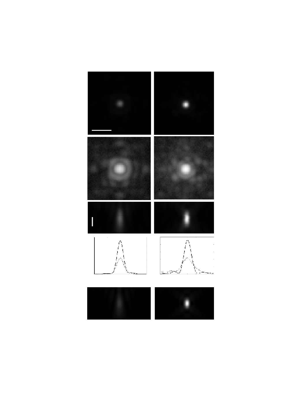
Figure. 15.12. shows. the. correction. of. a. 200. nm. bead. 67.μm. below. the. coverslip. in. a. glycerol/water.
mixture.with.a.refractive.index.of.1.42..Images.taken.irst.without.correcting.for.the.depth.aberration.
(a)
(b)
(c)
(d)
(e)
(f )
10
10
(g)
(h)
8
6
4
2
8
6
4
2
0
0
-
1.0
0.0
Lateral position (
μ
m)
-
0.5
0.5
1.0
-
3
-
2
-
1
0
1
2
3
Axial position (
μ
m)
(i)
(j)
FIGuRE 15.12
Images.of.a.200.nm.bead.67.μm.below.the.coverslip.in.a.water/glycerol.mixture.with.
n
.=.1.42..(a).
Uncorrected.image.of.in-focus.plane..(b).Corrected.image.of.in-focus.plane:.same.scale.as.(a)..(c.and.d).he.same.as.
(a).and.(b),.respectively,.but.on.a.logarithmic.scale..(e.and.f).Cross.sections.through.the.focal.plane.on.a.linear.scale..
he.scale.bars.are.1.μm..(g.and.h).Line.proiles.through.of.the.intensity.through.the.center.of.the.bead.along.a.lateral.
and.the.longitudinal.axis,.respectively..he.dashed.line.is.from.the.uncorrected.image.and.the.solid.line.is.from.the.
corrected.image..(i.and.j).Simulations.of.the.PSF..(i).Uncorrected.PSF.65.microns.into.a.material.with.index.1.42.
using.a.1.2.numerical.aperture.(NA).objective.with.a.1.512.refractive-index.immersion.oil..(j).A.simulated.PSF.at.
the.coverslip..he.peak.intensity.for.(j).is.3.75.times.the.peak.intensity.for.(i).(Kner.et.al..2010).