Biomedical Engineering Reference
In-Depth Information
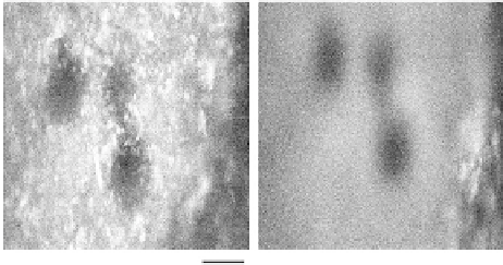
AO correction on microsphere fluorescence signal at 280
µ
m
AO on
AO off
50
µ
m
FIGuRE 12.17
he.microsphere.luorescence.signal.images.from.280.μm.below.the.surface.in.the.mouse.skull.
bone.with.AO.on/of.
To.create.the.luorescence.signal,.a.single.piece.of.mouse.skull.bone.was.bathed.in.the.green.luores-
cent. microspheres. solution. (0.1.μm,. Duke. Scientiic. Corp,. CA). for. 30. minutes.. his. process. allows. a.
large.number.of.microsphere.beads.to.penetrate.into.the.bone.cavity.and.act.as.the.luorescence.agents..
It.was.discovered.that.these.microspheres.located.in.the.deep.layer.are.capable.of.generating.enough.lu-
orescence.signal.for.imaging.as.well.as.for.AO.feedback..he.AO.correction.shows.tremendous.improve-
ment.in.the.quality.of.the.images..he.clusters.of.microspheres.can.be.resolved.with.the.AO.correction,.
as.shown.in.Figure.12.17.
With.increasing.imaging.depth,.the.high-frequency.aberrations—for.example,.the.second.spherical.
aberrations—become.signiicant..To.further.improve.from.the.current.AO.system,.a.woofer-tweeter.AO.
approach,.used.in.retinal.imaging.aberration.correction.[48],.can.potentially.be.used.to.improve.the.AO.
correction. in. the. two-photon. luorescence. imaging.. he. optical. aberrations. are. compensated. by. two.
deformable.mirrors..he.compensations.are.split.based.on.their.frequency.content..A.dedicated.DM.is.
used.to.correct.the.high-frequency.aberrations,.while.a.separate.DM.is.used.to.correct.the.low-frequency.
aberrations.
he.newly.developed.open-loop.DM.control.provides.another.potential.improvement.for.the.future.
development.of.the.sensorless.AO.[49]..he.DM.is.able.to.create.Zernike.shapes.in.difraction-limited.
precision.in.open.loop,.which.will.in.turn.improve.the.overall.residual.errors.of.the.SPGD-based.closed-
loop.AO.
12.7 Summary
he.BU-Wellman.Center.for.Photomedicine.AO.TPFM.is.used.to.correct.optical.aberrations.to.extend.
the.imaging.depth.in.mouse.bone-marrow.imaging..he.instrument.that.has.been.developed.includes.
a.Ti-sapphire.pulsed.laser,.a.MEMS.DM,.a.raster.scanning.system,.and.a.PMT.as.core.components..he.
SPGD.algorithm-based.AO.uses.the.luorescence.intensity.as.a.feedback.signal.to.drive.the.DM.to.com-
pensate.for.the.optical.aberrations..To.improve.the.AO.loop.eiciency.for.the.single-input-multi-output.
system,.a.Zernike.polynomial.basis.is.used.to.control.the.DM..he.Zernike.polynomial-based.AO.can.
compensate.and.enhance.the.microscope's.performance..he.PSF.of.the.microscope.was.measured.by.
using.a.simpliied.optical.coniguration.with.a.single.scanning.mirror..A.single.microsphere.luorescent.
bead.was.used.to.measure.the.PSF..Both.the.lateral.and.the.axial.resolutions.were.found.to.be.improved.
with.the.AO.correction.of.system.aberrations..he.lateral.and.the.axial.PSF.FWHMs.were.improved.17%.
and.31%,.respectively..AO.was.applied.to.deep.imaging.of.mouse.skull.tissue.in.a.genetically.modiied.
GFP.mouse..he.luorescence.signal.was.improved.81%.at.a.depth.of.145.μm.below.the.surface.from.AO.
correction..he.bone.cavity.structure.was.clearly.resolved.with.AO.at.a.depth.of.184.μm..Deeper.correc-
tion.was.explored.using.microspheres.as.luorescence.agents.in.the.mouse.skull.bone.