Biomedical Engineering Reference
In-Depth Information
mirror.(Newport.Corp.,.CA).capable.of.two-dimensional.scans.is.used.to.replace.the.polygon.and.the.
galvanometer.. he.main. reason. for. this.change. is.that.the.adjustable.scanning. angle. from. the.tip-tilt.
mirror.can.create.a.relatively.small.scanning.ield.with.suicient.pixel.resolution..For.example,.in.the.
setup.of.
Figure.12.9
,
.the.constructed.scanning.mechanics.is.able.to.provide.a.5.×.5.μm.scanning.ield,.
and.a.data.acquisition.board.(National.Instruments,.TX).is.able.to.grab.a.single.frame.at.50.×.50.pixels.
per.second..he.resulting.pixel.resolution.is.0.1.μm/pixel..To.achieve.a.three-dimensional.measurement,.
the. axial. direction. movement. is. controlled. by. a. linear. translation. stage. (Sutter. Instrument,. CA).. he.
stage.is.able.to.achieve.0.2.μm.resolution.
he. luorescence. agents. used. are. green. luorescent. microspheres. (Duke. Scientiic. Corp.,. CA).. he.
microsphere.has.a.diameter.of.0.1.μm.and.can.be.excited.at.920.nm.with.the.two-photon.excitation.pro-
cess..he.microspheres.are.diluted.in.water.with.agarose.powder.in.a.heated.environment.(85°C)..he.
cooled.inal.product,.agarose.gel,.is.used.as.the.imaging.target..he.agarose.gel.has.a.refractive.index.of.
1.33,.which.is.well.matched.to.the.water.immersion.objective.
Photobleaching.is.a.physical.process.that.will.damage.the.luorescence.agent.in.the.two-photon.excitation.
process..Consequently,.the.luorescence.signal.will.decrease.due.to.photobleaching..To.improve.the.signal-
to-noise.ratio.in.the.PSF.detection,.the.data.acquisition.(DAQ).is.running.at.a.speed.of.acquiring.1,000,000.
samples.every.second..Each.pixel.in.a.single.acquired.frame.will.be.the.average.value.of.400.samples.
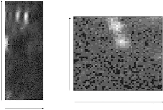
Figure.12.10.shows.the.measured.three-dimensional.intensity.distribution..To.identify.the.FWHM.
the.in.
x
-
z
.plane,.a.MATLAB.program.was.used.to.detect.the.maximum.pixel.value.of.a.single.frame.in.
the.
x
-
y
.intensity.distribution.for.each.measured.axial.position..his.maximum.intensity.cross.section.
in.the.axial.direction.was.then.itted.to.a.Gaussian.function..he.peak.value.from.the.Gaussian.function.
also.indicates.the.position.of.the.focal.plane.of.the.measured.focal.volume..he.
x
-
y
.intensity.measure-
ment. of. the. focal. plane. presents. the. information. for. the. lateral. resolution.. A. centroiding. program. is.
used.to.ind.the.center.of.the.mass.of.the.
x
-
y
.intensity.measurement.at.the.focal.plane,.and.Gaussian.
functions.were.it.to.the.one-dimensional.intensity.measurements.
he.measured.system.aberrations.are.dominated.by.low-order.aberrations:.astigmatism,.trefoil,.and.
coma..A.set.of.ive.PSF.measurements.are.shown.in.
Figure.12.11
..he.PSF.FWHM.averages.are.0.52.μm.
(lateral).and.3.09.μm.(axial).without.AO,.and.the.improved.PSF.FWHM.averages.are.0.43.μm.(lateral).
and.2.12.μm.(axial)..he.lateral.and.axial.PSF.FWHMs.are.improved.17%.and.31%,.respectively.
XZ plane
XY plane
0
µ
m
0
µ
m
5
µ
m
5
µ
m
FIGuRE 12.10
Images.of.luorescent.microspheres..Let,.the.luorescent.microspheres.in.the.
xz
.plane..Right,.he.
luorescent.microspheres.imaged.in.the.
xy
.plane.