Biomedical Engineering Reference
In-Depth Information
(a)
(b)
Circ-THG
Exc.
Lin-THG
Exc.
Lin-THG
Circ-THG
FIgurE 3.19
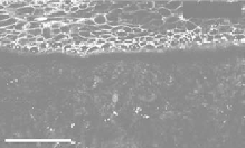
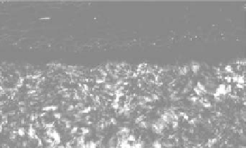
Influence of the excitation polarization on THG contrast. (a) THG image of a human cornea
recorded with linear incident polarization. (b) THG image of the same area recorded with circular incident polar-
ization. Scale bar = 50 μm. Conditions:
NA
= 0.8, λ = 1.2 μm. (Reprinted from Olivier N et al. 2010. Harmonic
microscopy of isotropic and anisotropic microstructure of the human cornea.
Opt. Express
18:5028-5040. With
permission of Optical Society of America.)
signals, since the epithelium is made of isotropic media. In contrast, the image from the same area
recorded with linear incident polarization also reveals the cellular structures with strong contrast.
In conclusion, the case of the cornea is an interesting illustration of the benefit of multimodal, non-
linear imaging: (i) THG and SHG images reveal different levels of organization of the microarchitecture
of the corneal stroma; and (ii) Polarization can be used to detect anisotropy.
3.4 conclusion
THG is a nonlinear imaging modality that uses a single excitation laser, so that it can be combined with
SHG and 2PEF microscopy. The contrast mechanisms of THG and SHG are different, so that the two
modalities provide different and complementary information. While SHG reveals a few number of orga-
nized structures with high specificity (fibrillar collagen, myofilaments, starch, astroglial fibers, polar-
ized microtubule assemblies, etc.), THG microscopy reveals optical heterogeneities in a general manner.
In cells, strong THG signals are observed from dense lipidic, mineralized, or absorbing structures. At
the tissue scale, THG imaging provides rich 3D morphological information. One remarkable property
of THG microscopy is that no signal is obtained from homogeneous media, implying that heterogene-
ities and interfaces are revealed with a good contrast. THG is particularly sensitive to structure size in
the 100 nm−2 μm range.
THG imaging has been reported in a number of contexts including imaging developing embryos
[37,39] brain tissue [26], neurons [10], hamster oral cavity [25], human skin [52], elastic fibers [53], lipid
droplets [27], hemozoin pigments [35], and so on. THG from organized biological media have been
reported from calcite [44], tooth dentin [54], and the human cornea ([24,47]). While few “real” applica-
tions have been reported to date (one example being the reconstruction of the early zebrafish develop-
ment [21]), it is anticipated that THG microscopy will find many uses as an “add-on” imaging technique,
owing to its versatility and its relatively straightforward combination with 2PEF−SHG microscopy.
Acknowledgments
We thank Marie-Claire Schanne-Klein for many discussions and comments, Maxwell Zimmerley for
critical reading, and Jean-Louis Martin for constant encouragement. We are indebted to all the colleagues