Biomedical Engineering Reference
In-Depth Information
(a)
(b)
3
B-THG
F-THG
2
B-THG
F-THG
1
δe
0
0.1
1
Axial period (µm)
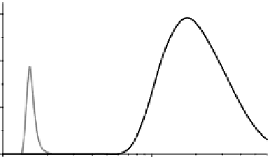
FIgurE 3.17
Quasi-phase matching. (a) Geometry considered: the medium exhibits a sinusoidal variation of
χ
(3)
along the optical axis. (b) Calculated F-THG and B-THG signal as a function of the axial period of the sam-
ple. (Reprinted from Débarre D, Olivier N, Beaurepaire E 2007. Signal epidetection in third-harmonic genera-
tion microscopy of turbid media.
Opt. Express
15:8913-8924; Olivier N, Beaurepaire E 2008. Third-harmonic
generation microscopy with focus-engineered beams: A numerical study.
Opt. Express
16:14703-14715. With
permission of Optical Society of America.)
number of emitters within the focal volume. This allows a direct comparison of the THG signal that is only
due to the quasi-phase matching. In both cases, we notice a large enhancement of the harmonic signal for
a particular sample periodicity. The theoretical value for the QPM resonance period can be expressed as
δe
= 2.
l
c
(3.28)
It can be noted that the B-THG resonant period corresponds closely to the theoretical value
(
l
c
BTHG
≈ 70nm and the maximum B-THG is obtained for δ
e
≈ 150 nm), as the phase and intensity dis-
tribution can be approximated as constant at this scale.
While this geometry demonstrates that more complex phase-matching conditions can be obtained in
organized media, it describes a hypothetical situation, since most organized media also have an orga-
nized microstructure which means that they exhibit some degree of anisotropy and tensorial aspects
have to be taken into account.
3.3.5 cornea imaging
In this section, we discuss combined THG−SHG of the human cornea. We show how multimodal non-
linear imaging can provide multiscale complementary information about the structure of this highly
organized biological medium.
3.3.5.1 Structure of the cornea
The outermost part of the cornea is an epithelium consisting of 6-7 cell layers. Beneath this epithelium
is a 500 μm-thick collagenous stroma. The stroma consists of layered 2 μm-thick collagen lamellae with
alternate orientations. These lamellae themselves are made of aligned collagen fibrils.
3.3.5.2 tHG and SHG Signals in the cornea
An example of combined THG−SHG imaging is shown in Figure 3.18. Multiharmonic images provide
a rich description of the lamellar organization of the intact stroma over its entire thickness. One inter-
esting observation is that THG and SHG signals are very different and generally exhibit anticorrelated
maxima, as illustrated in Figure 3.18c.