Biomedical Engineering Reference
In-Depth Information
More generally, the rule is that the orientation visibility ratio is large for structures that are small
compared to the beam focus (see, e.g., Figure 3.9 in the case
NA
= 0.8). Calculations show that the ori-
entation response becomes isotropic for objects larger than ≈ 500 nm using
NA
= 1.2, and for objects
larger than ≈ 1.2 μm using
NA
= 0.8.
Above this “threshold,” structures are equally visible in both orientations (although resolution is aniso-
tropic); this is illustrated in Figure 3.9 in the case of the epithelium of the cornea. In contrast, below this
size threshold, visibility strongly depends on object orientation, as illustrated in Figure 3.9 in the case of
the yolk of the zebrafish embryo, where only axially oriented structures are visible on the
xz
reprojection.
3.2.10 epidetection
As outlined in the previous sections, THG from dielectric volumes is predominantly forward directed.
This is because of the large phase mismatch for backward radiation, preventing efficient phase matching
for counter-propagating THG. This implies that THG microscopy is exclusively a transmitted-detection
technique in transparent media.
However, when imaging a thick biological tissue, although coherent THG scattering initially occurs in
the forward direction, incoherent scattering by the tissue can redirect some of the harmonic light toward
the excitation objective. Since scattering of visible light by tissues is mostly forward directed, harmonic
photons need to travel several scattering mean free paths in the tissue without being reabsorbed before
they can travel back to the surface (see Figure 3.10). Epidetected THG imaging is therefore usually pos-
sible in scattering, weakly absorbing tissues [19], as illustrated in skin [25] and brain tissue [26].
Another possibility for epidetection is the detection of THG from efficient nano-sized volumes such
as nanoparticles or metal surfaces [19,28], from which the THG radiation pattern is nearly dipolar, and
therefore nearly as efficient in the forward and backward directions. Significant backward emission can
also arise from periodically structured objects as will be discussed in Section 3.3.4.
(a)
Objective
Sample
(c)
Epi-THG
Trans-THG
Scattering sample
(b)
50 µm
Epi-THG
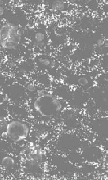
FIgurE 3.10
Epidetection of backscattered THG light. (a) In transparent samples, THG is co-propagating with
the excitation beam and is most efficiently detected in transmission. (b) In thick scattering samples such as biologi-
cal tissues, scattering can redirect a fraction of the THG light toward the excitation objective, so that epidetection is
possible. (c) Epidetected THG image of rat lung tissue. (Reprinted by permission from Macmillan Publishers Ltd.
Nat. Methods.
Debarre D et al. Imaging lipid bodies in cells and tissues using third-harmonic generation microscopy.
3:47-53. Copyright 2006.)