Biomedical Engineering Reference
In-Depth Information
b
(a)
(b)
(c)
Collagen remnants
d
100 μm
100 μm
c
(d)
(e)
200 μm
100 μm
100 μm
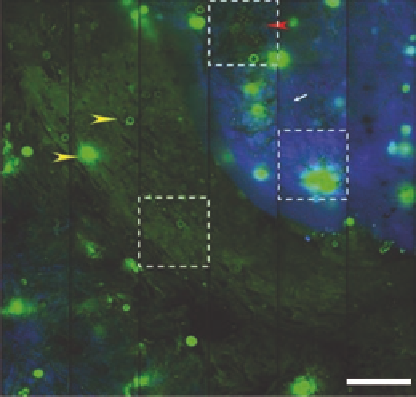
Figure 12.4
(a) Large-area, multiphoton autofluorescence (green) and backward SHG (blue) images of the
ex vivo
human cornea that have been infected with
Acanthamoeba castellinii
and
Pseudomonas aeruginosa
. Intrinsic autofluo-
rescence shows the presence of
Acanthamoeba
cysts (yellow arrowhead) and
Pseudomonas bacteria
(red arrowhead)
while backward SHG signal shows the degradation of corneal collagen. Increase in autofluorescence was also observed
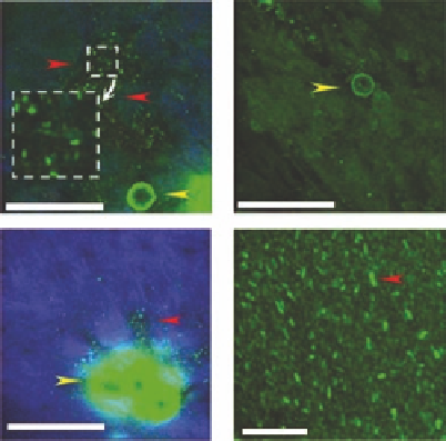
in infected cornea. (b), (c), (d) Magnified images from the selected regions of interests in (a). The presence of patho-
gens can be clearly visualized from autofluorescence. (e) Multiphoton autofluorescence image of isolated
Pseudomonas
aeruginosa.
(Adapted from Tan, H. Y. et al. 2007.
J Biomed Opt
12:024013.)
b
(a)
(b)
(c)
d
30 μm
30 μm
c
(d)
(e)
Segmentation
Green: fluorescence
Blue: SHG
Bifurcation
200 μm
30 μm
20 μm
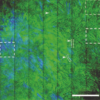
Figure 12.5
(a) Large-area, multiphoton autofluorescence (green) and backward SHG (blue) images of the
ex vivo
human cornea that have been infected with
Alternaria
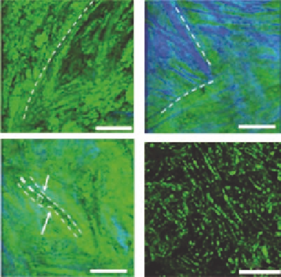
sp. (b), (c), and (d) represent the magnified areas b, c,
and d in (a). Parallel distributed fluorescence in the background and residual SHG-generating collagen remnants
were found in (b) and (c). Possible fungal hyphae with characteristic morphology of bifurcation and segmentation
(white arrows) can be visualized both in the large-scale (a) and detailed image (d). (e) Multiphoton autofluorescence
image of isolated
Alternaria
sp. (Adapted from Tan, H. Y. et al. 2007.
J Biomed Opt
12:024013.)