Biomedical Engineering Reference
In-Depth Information
16.2 imaging Methods
16.2.1 nonlinear imaging techniques for collagen characterization
Nonlinear optical imaging techniques, such as multiphoton laser scanning microscopy (MPLSM),
[29] are particularly useful for studying the changes taking place at the tumor−stroma boundary [30].
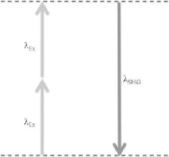
MPLSM is an optical sectioning technique that can simultaneously produce multiphoton excitation
(MPE) and second-harmonic generation (SHG). MPE occurs when two or more low-energy photons
excite a fluorophore, which then emits a single photon with higher energy than the individual incident
photons [29,31] (Figure 16.3). This method restricts fluorophore excitation to the plane of focus (i.e.,
optical sectioning) and reduces phototoxic effects [30,32] while increasing the effective imaging depth
in comparison to conventional confocal microscopy [33].
The noncentrosymmetric crystalline structure of fibrillar collagen I makes it an ideal candidate for
imaging via SHG microscopy [34,35]. SHG is a coherent, nonlinear, second-order polarization result-
ing from the nonabsorptive interaction between a pulsed laser source and a medium lacking a center of
symmetry [30,36]. Other key advantages of SHG involve optical sectioning and lack of photobleaching.
Additionally, if both forward and backward SHG is collected, information about the structure of col-
lagen can be inferred.
In addition to the collection of SHG, MPLSM is also useful for imaging a number of endogenous
(ex: NADH, FAD) and exogenous (ex: GFP, phalloidin) fluorophores. Imaging of fluorophores can be
used to determine the metabolic state of a cell or as markers for a range of molecular events within a cell.
This information can then be combined with SHG data to create a complete picture of collagen structure
and the states of associated cells.
Using SHG, collagen has been exploited as an optical biomarker in research. Being a fundamental
component of the cellular microenvironment, collagen has been visualized to study normal and abnor-
mal cellular behavior. As detailed above, Provenzano et al. (2006) [17] used this approach to describe
tumor associated collagen signatures (TACS) and the corresponding changes in collagen as breast can-
cer progresses. More recently, collagen order and ovarian cancer were investigated with forward and
backward scatter collection of SHG [37] to further work done by Kirkpatrick et al. [38].
16.2.2 SHG versus Gold Standard Histology Approaches
In addition to SHG, collagen can be visualized by staining with picrosirius red, a histology stain pop-
ularized in the 1980s. Initially Sirius red served as a substitute for acid fuchsin in Van Gieson's tri-
chrome to preserve the sample's colors for longer periods of time, up to months [39]. Trichromes, unlike
FIgurE 16.3
Jablonski diagram showing the two excitation photons and the single SHG emission photon.