Biomedical Engineering Reference
In-Depth Information
instead, the cellular and morphological information provided by THG modality plays a significant and
indispensible role in the diagnosis of the SSM.
14.3.4.4 Diseased Human Skin: Pigmented Basal cell carcinoma
BCC is the most common type of skin cancer and pigmented BCC is a variant of nodular BCC. Owing
to the abundant pigment in this lesion, it is sometimes misdiagnosed as malignant melanoma and an
in vivo
virtual biopsy tool with the ability to pathologically distinguish pigmented BCC and pigmented
nevi from melanoma is required. In histology studies of pigmented BCC (Agero et al. 2006), tightly
packed tumor cells bud from epidermis to papillary dermis with a nodular pattern enclosed by collagen
fibers. The nuclei of the tumor cells appear as a parallel arrangement (palisading) while melanin pig-
ment is nonuniformly distributed throughout the tumor nodules. Sometimes, a polarized cell pattern
can be seen in epidermis with loss of the normal progressive changes of keratinocytes, loss of the honey-
comb pattern, and loss of the dermal papillae structure. In previous researches (Cicchi et al. 2008), two-
photon imaging modality, combined both SHG and 2PF microscopy, has been applied to investigate
human
ex vivo
BCC samples, and the SHG/autofluorescence ratio of the dermis was found to be able to
discriminate between normal and BCC tissues. Figure 14.18 shows the
ex vivo
SHG/THG images of the
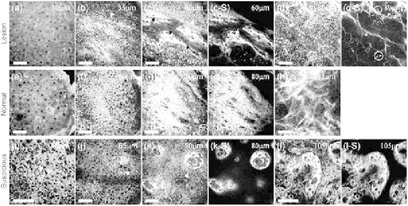
freshly excised pigmented BCC (Figures 14.18a through 14.18d), normal skin (Figures 14.18e through
14.18h), and suspicious tissue around the tumor (Figures 14.18i through 14.18l). At the dermo-epider-
mal junction, no dermal papilla architectures can be observed by SHG modality and the collagen fibers
at the junction are shown with a much wavier pattern (arrows in Figure 14.18c); this is in contrast to the
normal areolar pattern (Figure 14.18g). In contrast to the collagenous structures of the reticular dermis
observed in the normal skin (Figure 14.18h), the tumor nodules enclosed by the collagen fibers, which
FIgurE 14.18
Ex vivo
SHG/THG images of freshly excised (a)-(d) pigmented BCC specimen; (e)-(h) normal skin
specimen; and (i)-(l) suspicious cancerous or precancerous specimen obtained at different depths beneath the skin
surface. In lesional skin, the spindle-like keratinocytes (arrowheads in (b) and (c)) were usually found in epidermis
and collagen fibers in a much wavier pattern (arrows in (c) and (d)) can be found at the dermo-epidermal junction.
Without the normal pattern of rete ridges, tumor nodules (dashed arrow in (c)) enclosed by collagen fibers (arrow-
heads in (d)) were found to occupy the normal dermis. The parallel arrangement of the tumor cells (palisading; arrows
in (d)) can be identified at the edges of the nodules and bright HG spots (circled in (d)) can suggest the high melanin
contents. In the suspicious specimen, the spindle-like keratinocytes were observed in the epidermis (arrowheads in
(i)). Elongated basal cells can be found around the dermal papilla and were parallel to one another (arrowheads in (j)
and (k)), while (l) ring-shaped instead of (g) areolar collagen fibers were found in the papillary dermis. SHG images
corresponding to (c)-(d), (g), and (k)-(l) are shown in (c-S)-(d-S), (g-S), and (k-S)-(l-S), respectively. Scale bar: 50 μm.