Biomedical Engineering Reference
In-Depth Information
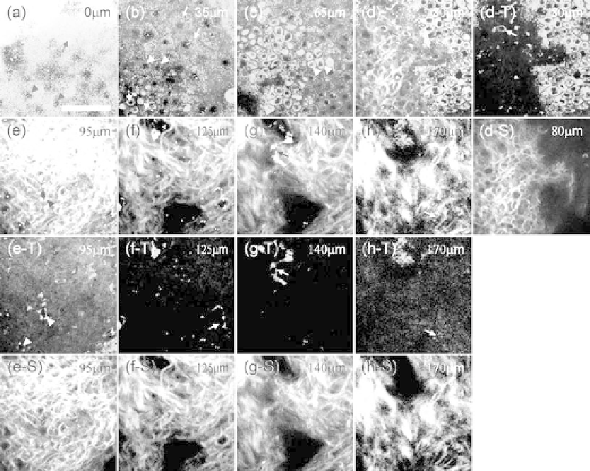
FIgurE 14.15
A series of SHG/THG images of the normal human skin obtained at (a) 0 μm, (b) 35 μm, (c)
65 μm, (d) 80 μm, (e) 95 μm, (f) 120 μm, (g) 140 μm, and (h) 170 μm beneath the skin surface. In (a), the stra-
tum corneum (arrow) with ultrastrong THG contrast and THG-dark nuclei of the granular cells (arrowheads)
are shown. (b) shows the granular cells (arrows), spinous cells (arrowheads), and basal cells (dashed arrows). (c)
The THG-brighter basal cells (arrowheads) covering the peak of the dermal papilla. (d) At the dermo-epidermal
junction, the collagen fibers in the peak of the dermal papilla surrounded by the basal cells are shown by SHG
microscopy and the inactivated melanocyte (arrow) can be found in the dermis. (e) Fibroblasts (arrowheads) can
be observed within the collagen fibers in the papillary dermis. (f)-(h) The collagenous structures of the reticular
dermis and the fibroblasts (arrows) within the fibers. THG and SHG images corresponding to (d)-(h) are shown in
(d-T)-(h-T) and (d-S)-(h-S), respectively. Scale bar: 50 μm.
by the interfaces of the multilayered structure (Tsang 1995; Barad et al. 1997) and by lipids (Debarre
et al. 2006) within the corneocytes. Based on the THG contrast from cytoplasmic organelles (Hsieh
et al. 2008), the nuclei of the epidermal cells appear dark (arrowheads in Figure 14.15a) in contrast to the
bright cytoplasm in the THG images. Through THG modality, all the granular cells (arrows in Figure
14.15b), spinous cells (arrowheads in Figure 14.15b), and basal cells (dashed arrows in Figure 14.15b) can
be found with honeycomb architectures throughout all levels of the epidermis. The progressive changes
of nuclear diameter, cell diameter, and cell density in different layers can be clearly revealed to indicate
the keratinization process. In the stratum basale (Figure 14.15c), clusters of basal cells (arrowheads)
show stronger THG contrast relative to the surrounding cells and cover the peak of the dermal papilla
at the “bumpy” dermo-epidermal junction. These strong THG contrasts in the cytoplasm of the basal
cells are found to strongly correlate with the high concentration of the melanin in the basal cells and are
identified to be contributed by the absorption resonance enhancement of the melanin (Chen et al. 2010).