Biomedical Engineering Reference
In-Depth Information
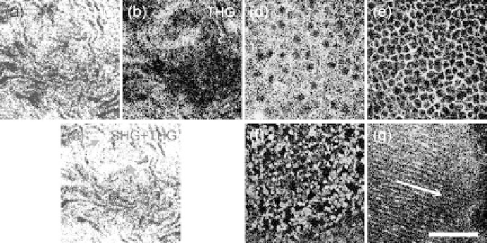
FIgurE 14.14
SHG/THG images of an excised mouse eye obtained at different depths beneath the corneal sur-
face. (a) and (b) show the SHG and THG images of the corneal stroma and (c) shows the corresponding combined
SHG/THG image. The collagenous structures can be revealed by SHG, while the keratocytes lying within the colla-
gen fibers can be recognized from the non-SHG-overlapped THG signals (arrows). Through THG modality, the cel-
lular structures of the (d) upper corneal epithelium, (e) deeper corneal epithelium, and (f) corneal endothelium can
be revealed, while (g) the lens fibers in the lens are shown clearly along the direction of the arrow. Scale bar: 50 μm
.
14.14c), THG and SHG are found to overlap in some regions (shown white in Figure 14.14c) but do not
overlap in other regions. Since THG contrast can also be provided by the optical inhomogeneity of the
collagen fibers, the SHG-overlapped THG signals can be recognized to arise from the collagen fibers
(shown white in Figure 14.14c). On the other hand, the non-SHG-overlapped THG signals reflect the
keratocytes lying within the collagen fiber meshes (arrows in Figure 14.14c). By combining SHG with
THG modality, not only can the collagenous structures be observed, but also the cellular information in
the CS can be distinguished and obtained more easily.
In addition to the CS, the outer and inner cellular layers, EP and ED, also play important roles in
ophthalmology. ED covers the anterior surface of the cornea and has five to six cell layers thick, while
the ED is a monolayer of flattened and polygonal ED cells. The EP cells of the superficial layer (close to
the anterior surface of the cornea) are squamous and the EP cells of deeper layers are columnar. The EP
and ED are responsible for protection and governing the fluid transportation of the eye, respectively.
For diagnosis of many eye diseases, to highly resolve the cell morphology of these two layers is quite
significant and an imaging tool with high sectioning power is required. Based on the THG contrast aris-
ing from the cytoplasmic organelles (Hsieh et al. 2008), the cytoplasm of the EP cells appeared bright
in contrast to the dark nuclei. By optically sectioning the cornea at different depths, the morphology of
the EP cells can be successfully revealed (Figures 14.14d through 14.14e) to be consistent with the histol-
ogy results (Ramaesh et al. 2004). From the superficial layer (Figure 14.14d) to the deeper layer (Figure
14.14e), the shape of the EP cells is found to change from squamous shape to columnar shape. Figure
14.14f shows the THG image of the ED at 120 μm, and the uniformly sized polygonal cells in this mono-
layer could be revealed. The nuclei of the endothelial cells appear dark (arrowheads in Figure 14.14f),
while the cytoplasm appears bright. Beneath the cornea is the aqueous humor (AH), which is mainly
composed of water and has no THG contrast due to its optical homogeneity. Passing through the AH is
the lens (L), consisting of three main parts: the membrane-like lens capsule (LC), cellular lens epithe-
lium (LE), and lens fibers (LF). With a high penetrability of greater than 700 μm (Chen et al. 2009b), at
a depth of 430 μm beneath the anterior corneal surface, the lens fibers (Figure 14.14g) with a width of
~5 μm (along the arrow in Figure 14.14g) can be highly resolved through THG signals but no significant
SHG signals can be observed from the lens fibers. Even at a depth of >700 μm, the structure of the lens