Biomedical Engineering Reference
In-Depth Information
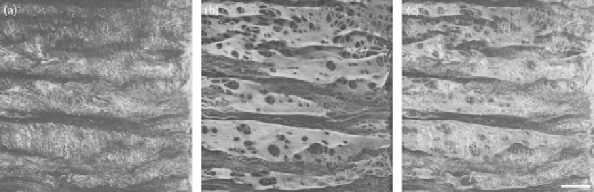
FIgurE 13.6
(
See color insert
.) Porcine renal artery wall.
En face
view of 3D reconstructed artery. (a) Collagen
SHG. (b) Elastin autofluorescence. (c) Merged image. Scale bar = 50 μm.
Several investigators have used two-photon excited elastin autofluorescence to quantify various
physical characteristics of arterial elastic laminae. Megens et al. found the size (1.2 versus 2.1 μm) and
density (0.045 versus 0.57 μm
−2
) of internal elastic lamina fenestrae differed between murine elastic
(carotid) and muscular (uterine and mesenteric) arteries [32]. Kwon et al. found holes in surface elastin
in the porcine carotid artery with diameters that ranged in size from 3 μm (circular) to 25 × 9 μm (oval)
with a density of 0.002-0.004 μm
−2
[39].
13.4.2 collagen
Collagen fibers in the arterial wall provide tensile strength and thereby are a major determinant of
vascular integrity. In the many types of collagen, including the fibril-forming collagens Types I, II, III,
V, and XI, three protein chains align to form triple helical molecules. Secreted triple helical molecules
(~1.5 nm diameter and 300 nm long) spontaneously assemble into five-stranded microfibrils (~4.5 nm
diameter). The microfibers aggregate end to end and laterally in a radial pattern to form fibrils (10-
100 nm diameter), which then cluster to form much larger fibers (1-100 μm), that are arranged in large
bundles readily seen by light microscopy [43].
As mentioned in Section 13.2.1.2, based on its intrinsic properties, collagen can be imaged without
the need for fixation or staining. Collagen generates both two-photon excited fluorescence and sec-
ond harmonic photon emissions. Two-photon excitation of collagen at shorter excitation wavelengths
(<800 nm) can generate both SHG and two-photon excited fluorescence (with a weak and broad spec-
trum), whereas at higher excitation wavelengths, the collagen signal is due to SHG alone [44].
13.4.3 Macromolecular exogenous Probes
Exogenous fluorescent probes provide an alternate means to detect arterial wall macromolecu-
lar microstructures by two-photon excitation microscopy. The collagen-binding adhesion protein
35-Oregon Green 488 (CNA35-OG488) is a fluorescently labeled molecular imaging agent developed
for optical imaging of collagen [43]. CNA binds to fibrillar collagen types I, III, and IV [43], all of
which are abundant in the arterial wall, and are associated with atherosclerosis and plaque progres-
sion. Studies using CNA have provided insights into the sites where diffusion barriers are present in
the arterial wall. In healthy muscular arteries, all layers are labeled e
x vivo
; however, in elastic arter-
ies, medial and intimal labeling appears to be prevented by endothelium and elastic laminae [45].
After mechanical damage to the carotid artery, and in blood vessels with discontinuous endothelial
vascular coverage (liver, spleen), or fenestrated endothelium (kidney), subendothelial collagen was
strongly labeled but not in vessels in organs with continuous endothelium (heart, lungs). This finding