Biomedical Engineering Reference
In-Depth Information
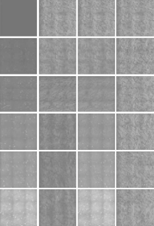
TPAF
glycated
SHG
glycated
Composite
glycated
Composite
control
Day 0
Day 2
Day 4
Day 6
Day 8
Day 10
FIgurE 12.10
(
See color insert.
) Time-coursed multiphoton autofluorescence (green) and SHG (red) images of gly-
cated and control (nonglycated) samples of bovine cornea. Frame size is 620 × 620 mm
2
. (Adapted from Tseng, J. Y.
et al. 2011.
Biomed Opt Express
2(2):218−230.)
fiber in the formation of autofluorescent AGEs, which may be useful for early detection of diabetes
mellitus-induced ophthalmological pathologies.
As a final example of the ability of multiphoton imaging in visualizing pathological corneas, bovine
corneas were immersed in de-ionized water for 2 h to simulate cornea edema [69]. Following induced
edematous conditions, the corneas were mounted for FWSHG and BWSHG imaging. Shown in Figures
12.12a 12.12b, 12.12e, and 12.12f are FWSHG and BWSHG image stacks of untreated bovine corneas.
On the other hand, Figures 12.12c, 12.12d, 12.12g, and 12.12h represent image stacks from edematous
corneas. In both cases, SHG images were acquired from both the anterior and posterior stroma. Note
that in edematous corneas, lamella oriented obliquely to the corneal surface (arrowheads in Figure
12.12d) and increased lamellae spacing (arrowheads in Figure 12.12h) can be visualized.
Enlarged
en face
forward SHG (FWSHG) and backward SHG (BWSHG) images of untreated and
edematous bovine corneas at depths of 100 and 1000 μm were illustrated in Figure 12.13. As noted in the

