Biomedical Engineering Reference
In-Depth Information
(a)
(c)
I
III
IV
III-1
I-1
II
III
I
Parallel aligned collagen bundles
400 μm
400 μm
V
I-1
III-1
1000 μm
(b)
Elongated epithelial cells
Parallel aligned collagen bundles
100 μm
100 μm
FIgurE 12.6
(a) Large-area, multiphoton autofluorescence and backward SHG images of the
ex vivo
human
keratoconical cornea. Intrinsic autofluorescence shows the presence of the keratoconical apex while backward SHG
signal identified the global organization of collagen fibers around the apex. (b) Corneal topography shows that the
location of the keratoconical apex is consistent with that found from multiphoton imaging. (c) I and III are magni-
fied images from the selected regions of interests in (a). Further enlargement of the images show that epithelial cells
near that apex are elongated (I-1) and that backward SHG patterns near the apex are parallel aligned. (Adapted from
Tan, H. Y. et al. 2006.
Invest Ophthalmol Vis Sci
47:5251-5259.)
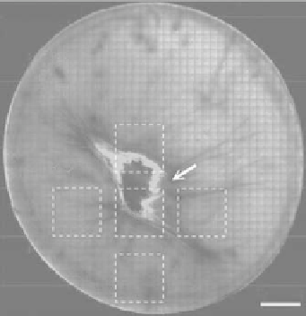
Physical trauma to the cornea represents another condition that multiphoton imaging can be useful
for diagnostic purposes. Shown in Figure 12.7 are large-area, multiphoton autofluorescence and BWSHG
images of an excised human cornea that is known to contain a scar. Near the corneal surface (0 μm), cor-
neal epithelium can be identified by its intrinsic autofluorescence, and backward SHG signal illustrates
corneal collagen protruding across the Bowman layer. At the depth of approximately 1200 μm, wound
regions lacking SHG signal were observed. Along the wound edge, intense autofluorescence and BWSHG
pattern aligned in parallel to the wound edge were both observed. The ability of multiphoton microscopy
identifying the structural alteration of the cornea is again demonstrated.
In addition to diagnosing corneal pathologies of human cornea specimens, multiphoton microscopy
may also be used to study the processes associated with corneal pathologies under controlled conditions.
One experiment that we performed was to simulate infection caused by
Pseudomonas aeruginosa
in
bovine corneas
in vitro
[14]. Following artificial injection of the pathogen into the cornea, the specimens
were kept at 37°C and multiphoton images were acquired at fixed intervals following pathogen injec-
tion. In this manner, we were able to follow the temporal effect of pathogen infection in corneas. Shown
in Figure 12.8 are our results, large-area, multiphoton autofluorescence and BWSHG images of excised