Biomedical Engineering Reference
In-Depth Information
Corneal
epithelium
Descemet's membrane
Collagen
Corneal collagen
100 μm
0 μm
862 μm
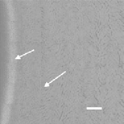
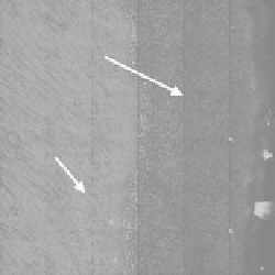
FIgurE 12.2
Large-area, multiphoton autofluorescence and backward SHG images of the
ex vivo
porcine cornea
at different depths. At the corneal surface (0 mm), corneal epithelium can be identified by its intrinsic autofluores-
cence while backward SHG signal from stromal collagen can be used for imaging purposes. At the greater depth
of approximate 862 mm, autofluorescence can be used to characterize the Descemet's membrane and endothelial
region while backward SHG can be detected at the deepest layer of stromal collagen. (Adapted from Teng, S. W. et
al. 2006.
Invest Ophthalmol Vis Sci
47:1216-1224.)
used to characterize the limbal cells. The ability of acquiring limbus structure information and its
biochemical state is of considerable significance because of the proliferation capability of the stem
cell within the limbus and its potential application in tissue engineering. Unlike corneal collagen,
BWSHG shows that scleral collagen is more randomly organized. In addition, owing to the shorter
coherent length of BWSHG radiation, the forward SHG (FWSHG) signal is usually stronger than
BWSHG and exhibits the fibrous morphology in corneal imaging better [61]. As a result, FWSHG has
been frequently used to investigate the corneal collagen fibril orientation [62,63]. Nonetheless, the
FWSHG signal in the clinical or
in vivo
observation is practically unavailable. Therefore, understand-
ing the nature of BWSHG is of considerable significance in both normal and pathological imaging of
the cornea.
Scleral collagen
Limbal epithelium
(melanin containing)
Cornea
collagen
100 μm
0 μm
50 μm
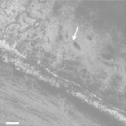
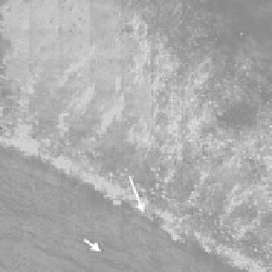
FIgurE 12.3
Large-area, multiphoton autofluorescence and backward SHG images of the
ex vivo
porcine cornea
at different depths. Near the surface of corneal−scleral junction (0 mm), conjunctival and limbal epithelium can be
identified by its intrinsic autofluorescence while backward SHG signal from corneal and scleral collagen can be used
for imaging purposes. At the greater depth of approximate 50 mm, autofluorescence can be used to characterize the
limbal cells between corneal and scleral collagen. Backward SHG shows that unlike corneal collagen, scleral collagen
is more randomly organized. (Adapted from Teng, S. W. et al. 2006.
Invest Ophthalmol Vis Sci
47:1216-1224.)