Biomedical Engineering Reference
In-Depth Information
PMT
Broad band
autofluorescence
435 nm~700 nm
E 435 LP
700 sp
PMT
SHG
380~400 nm
Dichroic
435 dcxr
HQ 390/20
Quarter-wave
plate
Half-wave
plate
Ti-sapphire
pulse laser
Dichroic
mirror
Wavelength
780 nm
Nikon
WI, 40X
Objective
Polarizer
XY scanner
Sample
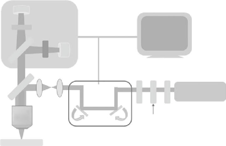
FIgurE 12.1
The experimental setup of multiphoton microscope.
the signals pass through the primary dichroic mirror. Next, the broadband multiphoton autofluores-
cence and SHG signals are separated by a secondary dichroic mirror (435dcxr, Chroma Technology)
and respectively filtered by two band-pass filters (MAF: E435lp-700sp, SHG: HQ390/20, Chroma
Technology) before reaching the detectors. For signal detection, photon-counting photomultiplier tubes
(R7400P, Hamamatsu, Japan) can be employed. The typical detection bandwidths of the broadband
fluorescence and SHG used in our laboratory are 435-700 and 380-400 nm, respectively.
12.3 Visualization of normal corneal and Surrounding tissues
In the pioneering work published by Tromberg's group, it was shown that SHG signal propagating in
the backward direction can be registered for visualization of corneal structures under
ex vivo
con-
ditions [57]. Our experimental results and that of others demonstrated the efficacy of multiphoton
microscopy for corneal imaging [13,59,60]. As shown in Figure 12.2, the large-area, multiphoton
autofluorescence and backward SHG images of the
ex vivo
porcine cornea were acquired at different
depths. At the corneal surface (0 μm), corneal epithelium can be identified by its intrinsic autofluo-
rescence originated from the cytoplasmic portion of cells and the lack of the signal from the nuclei.
Owing to the intrinsic curvature of the eye, backward SHG (BWSHG) signal from stromal collagen
can be observed at the same depth as the epithelium. At the greater depth of approximately 862 μm,
autofluorescence can be used to characterize the Descemet's membrane and endothelial region while
BWSHG can be detected at the deepest layer of stromal collagen. The BWSHG pattern reveals the
presence of the collagen fibers and indicates the difference of orientation between anterior and pos-
terior regions of the cornea. Other ocular components adjacent to the cornea, such as the limbus and
sclera, can also be effectively imaged by autofluorescence and SHG microscopy (Figure 12.3). Near
the surface of corneal-scleral junction (0 μm), conjunctival and limbal epithelium can be identified
by its intrinsic autofluorescence, and BWSHG signal emanated from corneal and scleral collagen can
be used for imaging purposes. At the greater depth of approximately 50 μm, autofluorescence can be