Biomedical Engineering Reference
In-Depth Information
(a)
(b)
(c)
(d)
3
2.5
2
1.5
1
0.5
0
0
5
pH 5.5
pH 6.5
4
3
2
1
0
TPF
e
/TPF
c
20
δ
(degrees)
40
60
80
SHG
e
/SHG
c
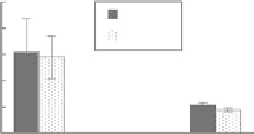
FIgurE 11.5
(a) Mask of in-plane-oriented collagen fiber cross-sections, taken from a TPF image of an acel-
lular collagen hydrogel. (b) Mask of out-of-plane-oriented collagen fiber cross-sections, taken from a TPF image
of an acellular collagen hydrogel. (c) The ratio of noise-subtracted SHG and TPF signal from the signal-containing
regions masked in (a) (elliptical particles,
e
) and (b) (circular particles,
c
). (d)
d
ef
2
, in arbitrary units, was estimated
for various angles of tilt of the collagen fiber axis with respect to the laser propagation direction. Fibers at 10° and
63° of tilt are diagrammed with respect to the laser focal region (gray). (Reprinted from
Biophys J.
, 94, Raub, C.
B. et al., Image correlation spectroscopy of multiphoton images correlates with collagen mechanical properties,
2361-2373, Copyright 2008, with permission from Elsevier.)
The 1:1 ratio of TPF signal from fibers predominantly parallel versus transverse to the image plane
confirms the isotropic angular distribution of TPF generation in collagen gels. In contrast, one would
expect greater backward-detected SHG from fibers parallel to the image plane, both because of scatter-
ing of forward-generated SHG at the fiber bottom interface [14], and because of the collagen dipoles'
optimal orientations with the laser propagation direction [73]. Indeed, a 3:1 ratio of SHG intensity from
fibers roughly parallel versus roughly transverse to the image plane was measured, which is consistent
with theoretical considerations using a lower numerical aperture (NA) [92]. A higher NA [1.3] objective
was used to collect the SHG and TPF images, introducing a component of the electric field transverse to
the image plane, which would change the SHG angular power distribution from fibers aligned with the
z
-axis. Specifically, these fibers should emit forward-generated SHG at a steeper angle (~40-47°) from
the
z
-axis compared to the lower NA case, but no backward-generated SHG due to the Guoy phase shift
and destructive interference{}. Fibers aligned parallel to the laser propagation should exhibit lower
d
ef
(
d
=
< ) as well as reduced single and multiple scattering of SHG photons from the highly forward-
directed SHG emission compared to fibers oriented perpendicular to the laser propagation direction.
Although a higher NA objective provides additional electric field contributions to SHG signal, the for-
ward-directed generation and lack of scattering interfaces in transverse collagen fiber sections could
explain the observed threefold difference in epi-detected SHG signal.
From Equation 11.5, a ratio of SHG signal intensities was calculated by estimating values of
d
ef
, which
depend on the orientation of collagen dipoles with respect to the incident electric field. This calculation
d
eff
eff


