Biomedical Engineering Reference
In-Depth Information
1.5 mg/mL
4.5 mg/mL
9 mg/mL
(a)
(c)
(b)
(e)
(f )
(d)
(g)
(h)
1.2
100
SHG coarse AF
SHG fine AF
1
0.8
0.6
0.4
0.2
0
0
SHG coarse
80
SHG fine
60
40
20
0
0.2 0.4
Normalized concentration
0.6
0.8
1
0
2
4
6
8
10
Collagen concentration (mg/ml)
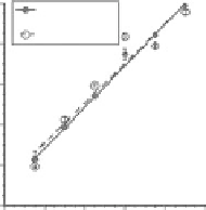
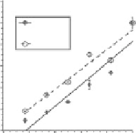
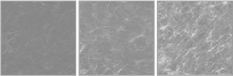
FIgurE 11.4
Simultaneously collected SHG and TPF (signals co-registered in grayscale) images of acellular col-
lagen hydrogels polymerized at pH 8.5 (a-c) or 14°C (d-f) and collagen concentrations of (a,d) 1.5 mg/mL, (b,e)
4.5 mg/mL, and (c,f) 9 mg/mL. (g) Normalized SHG signal versus normalized collagen concentration for the fine-
and coarse-structured gels, with linear best-fit lines shown. (h) SHG signal image area fraction versus collagen con-
centration for fine- and coarse-structured gels, with linear best-fit lines shown. The error bars represent standard
deviation. The data markers are indicated in the figure legend. The scale bar represents 50 μm.
11.3.5 effect of collagen Fiber orientation on SHG
Several studies have shown through theoretical and experimental methods that SHG depends upon
the orientation of collagen monomers aggregated into fibrils and fibers with respect to both the laser
polarization angle and the laser propagation direction [3,10-14,25,84,88]. Specifically, for a fibril per-
pendicular to the laser propagation direction, SHG is maximized when the dipoles within the fibril are
aligned parallel to the incident electric field and is minimized when the dipoles are perpendicular. The
coalignment of dipoles and the electric field allows for a maximum nonlinear polarization. This orienta-
tion dependence of SHG has been used to characterize the in-plane orientation of collagen fibrils since
it has been shown that the dipoles within collagen align with the fibril long axis [13].
The orientation dependence of SHG signal from collagen fibrils, though a useful structural parameter,
can interfere with accurate structural characterization of a collagen network from a single SHG image
or image stack since fibrils would possess variable SHG intensity depending upon fibril orientation. For
circularly polarized laser illumination, however, SHG signal does not depend upon fibril-dipole ori-
entation within the image plane [14]. However, there still exists an axial (out-of-plane) dependence, in
which SHG is maximized from fibrils perpendicular to the laser propagation direction (i.e., in the image
plane), and is minimized from fibrils parallel to the laser propagation direction (i.e., perpendicular to
the image plane). The following discussion addresses the axial dependence of SHG signal in collagen