Biomedical Engineering Reference
In-Depth Information
(a)
TPF (FM4-64)
SHG (FM4-64)
Alexa488
TPF
SHG
Alexa
50 µm
5 µm
(b)
12
10
8
6
10 mV
5 msec
0 mV
4
2
0
1 %
Soma
(
n
= 5)
Axon
(
n
= 5)
5 msec
0 %
6
5
4
3
2
(c)
10 mV
5 msec
0 mV
1%
1
0
5 msec
Soma
(
n
= 8)
Axon
(
n
= 11)
0 %
FIgurE 10.4
(
See color insert.
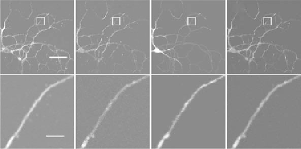
) Membrane potential measurements at axons. (a) Visualization of axons by SHG.
Primary cultured dissociated neurons were extracellularly stained with FM4-64 and loaded with Alexa 488 intra-
cellularly. The whole neuronal morphologies including fine processes of axons can be clearly visualized by SHG.
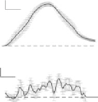
(b) Membrane potential changes at axons with action potential. The upper left panel shows the voltage changes
recorded at the soma and the lower left panel shows SHG changes simultaneously recorded at axons. Data are shown
as the mean (black) ± SEM (red) from five neurons. The right panel shows a comparison of the peak amplitude of
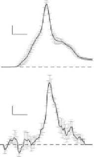
SHG signal changes normalized to the action potential with 100 mV amplitude. (c) Membrane potential changes
at axons with nonregenerating somatic voltage change. Left: Neurons were held under current-clamp conditions in
the presence of 1 μM TTX and injected with a depolarizing current pulse. The voltage change at the soma (upper left
panel) and corresponding changes of SHG recorded at axons (lower left panel) are shown. Data shown is the mean
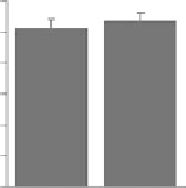
(black) ± SEM (red) from 12 recordings from 7 neurons. Right: Neurons were held under voltage-clamp configura-
tion and 50 mV voltage pulses were applied to the soma. SHG responses at the soma (
n
= 8 from 6 neurons) and
axons (
n
= 11 from 6 neurons) were measured. Data in the panel presented is mean ± SEM. (Adapted from Nuriya,
M. and M. Yasui. 2010.
J Biomed Opt
15:020503. With permission.)