Biomedical Engineering Reference
In-Depth Information
fibrils and interfibril spacing of the mature tendons were sufficiently large to produce predominantly
forward SHG and that the similar appearance in the backward channel may have been due to multiple
scattering of the initial
F
SHG
. This description can also be utilized to explain the observation of bright
B
SHG
from sclera by Han [6]. The hollow fibrils in this tissue are on the order of 300 nm and
a priori
would be expected to produce predominantly
F
SHG
. However, they reported
F
SHG
/B
SHG
~ 1. This result is
predicted by our current theory if one associates the domain length with the shell sidewall (presumably
much thinner than the diameter) rather than the fibril diameter.
6.4 Results of 3D imaging and Analysis
6.4.1 SHG imaging of the Murine Model of osteogenesis imperfecta
Having discussed our experimental methods, our simulation framework, and our heuristic model of SHG
production in tissues, we present the combined analysis for the murine oim model of osteogenesis imper-
fecta. This is a heritable disease of humans characterized by recurrent bone fractures, stunted growth,
defective teeth, and other symptoms from abnormal tissues composed of type I collagen. OI results from
mutations within the
Col1A1
or
Col1A2
genes that affect the primary structure of the collagen chain and
induce changes in the secondary structure of the collagen trimers, which incorporate the mutant chains.
The ultimate outcome is collagen fibrils that are either abnormally organized, small, or both.
6.4.1.1 Determination of the Bulk optical Parameters for oim and Wild type Skin
While the most dramatic clinical presentation of OI is in bone, we chose to examine skin. This tissue is
the most feasible for a virtual optical biopsy (i.e., backward collection geometry), and certainly be more
accessible than bone for
ex vivo
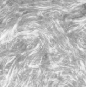
analysis. Representative single optical sections for the oim and wild
type (WT) skin are shown in Figure 6.5. While these tissues are distinct by visual inspection, where
the images suggest that the fibrils are less ordered for the diseased skin, for future clinical diagnosis, a
quantitative description is required. The less dense packing in the oim case suggests that the scattering
properties may be different. Here, we measured μ
s
and
g
(effective) values for the oim and WT skin at
both the fundamental and SHG wavelengths.
This scattering anisotropy,
g
, is a measure of the directionality of photon scattering, varies from 0 to
1, and is typically in the range of ~0.6-0.95 for most connective tissues. The upper limit corresponds to
highly forward-directed scattering and is characteristic of very highly ordered tissues such as tendon,
whereas in the other limit, brain is random and has low anisotropy. Thus,
g
can be used as a measure of
the organization of the tissue. The resulting
g
values for 900 and 457 nm are shown in Table 6.1. At the
SHG wavelength, fits of the experimental data to Equation 6.2 yield respective values for the oim and WT
(a)
(b)
FIgurE 6.5
SHG single optical sections of murine WT (a) and oim dermis (b). Scale bar = 25 microns. (Reprinted
from
Biophys. J
. 94, Lacomb, R., O. Nadiarnykh, and P. J. Campagnola, Quantitative SHG imaging of the diseased state
osteogenesis imperfecta: Experiment and simulation, 4504-4514, Copyright (2008), with permission from Elsevier.)


