Biomedical Engineering Reference
In-Depth Information
The underlying coherence of the SHG is the enabling factor in elucidating these structural details.
Incoherent modalities such as fluorescence and reflectance confocal cannot reveal this level of detail.
We note that while collagen does possess weakly imageable autofluorescence, the emission typically
appears somewhat diffuse and is not necessarily reflective of the fibrillar structure.
An example demonstrating how the coherence of SHG can uniquely obtain information imaging
collagen in diseased states comes from our group, where we compared the structure of normal and
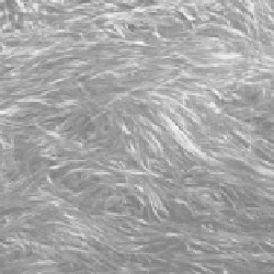
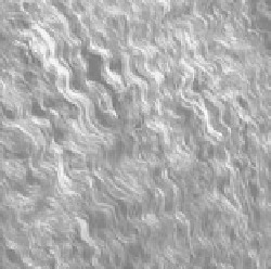
malignant ovarian tissues (almost all type I collagen). Sharp differences exist for collagen morphology
in these tissues [38], where the malignant collagen fibers display higher regularity and are more densely
packed along a predominant direction, while the normal collagen fibers are loosely distributed in all
directions (Figure 4.9). This change in morphology was also evident in the directional data, which,
along with Monte Carlo simulations, allowed us to deduce changes in the fibril size and distribution.
Concurrently, SHG polarization anisotropy revealed changes in the fiber morphology. More details on
this aspect are presented in Chapter 6. Thus, as is the case with cornea, the coherence enables subresolu-
tion information on the fibril assembly to be obtained.
Besides fibrillar collagen tissues, several other endogenous protein structures in biological sys-
tems have been investigated using SHG microscopy, where this modality produces unique contrast
relative to the other NLO techniques. Detailed features of actomyosin structures in muscle tissue
(a)
Normal ovary
Ovarian cancer
(b)
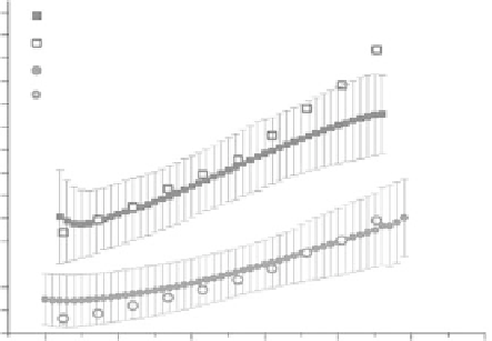
8.0
Normal, measured
Normal, simulated
Cancer, measured
Cancer, simulated
7.0
6.0
5.0
4.0
3.0
2.0
1.0
0
20
40
60
80
100
120
Depth (microns)
FIgurE 4.9
(a) Representative SHG images of collagen fibers in normal human ovary (left) and malignant ovary
(right) with 890 nm excitation wavelength (field size = 170 μm). (b) Forward/backward SHG ratio versus imaging
depth for normal and malignant ovarian tissues and Monte Carlo simulations using measured bulk optical param-
eters. (Adapted from Campagnola, P. 2011.
Anal. Chem
. 83:3224-3231.)