Biomedical Engineering Reference
In-Depth Information
O-P-O
Amide I
CH
H
2
O
1000
1500
2565
2575
1000
1500
2000
2500
3000
3500
cm
-1
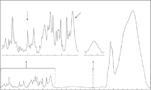
FIgurE 4.5
The Raman spectrum of the P22 virus showing characteristic vibrational frequencies observed
in biological samples. Several vibrational modes of particular interest in vibrational microscopy are labeled. The
O-P-O stretching vibration arises from the vibration of the DNA backbone. The amide-I band is characteristic of
proteins and can be used to map out protein density. The CH-stretching band is typically used to image lipids. The
H
2
O-stretching vibrations of water are important for following water flow and density. (Adapted from Evans, C. L.
and X. S. Xie. 2008.
Annu. Rev. Anal. Chem.
1:883-909. With permission.)
wavelength can be in the visible range, leading to higher spatial resolution. Indeed, confocal spontane-
ous Raman microscopy has been previously developed to provide a “chemical” imaging tool with 3-D
capability [29].
However, owing to the small scattering cross section (~10
−30
cm
2
compared to ~10
−16
cm
2
for absorp-
tion of a typical dye), the spontaneous Raman signal is weak. This is especially true for anti-Stokes
Raman scatter as it is usually much weaker than Stokes Raman scattering, because anti-Stokes Raman
scattering occurs from vibrationally excited molecules (Figure 4.4), whose population is many orders
of magnitude smaller than the molecules in the ground state. The relative population for molecules in
the vibrationally excited and ground states can be estimated from the Boltzmann distribution law by
~
e
−
ω
, where
ħ
ω
vib
is the energy gap between the two vibrational energy levels shown in Figure 4.4,
and
R
and
T
are the gas constant and the absolute temperature, respectively. For the CH-stretching
vibrational band that is typically chosen to image lipids,
ħ
ω
vib
is ~3000 cm
−1
. At room temperature,
RT corresponds to ~200 cm
−1
. From this, it can be calculated that only 0.3 ppm of lipid molecules is
in the CH-stretching vibrationally excited state. Apparently, to use the Stokes or anti-Stokes Raman
scattering signal for imaging of living specimens, where fast acquisition is required, strong enhance-
ment mechanisms such as CARS and SRS need to be implemented to increase the sensitivity by several
orders of magnitude.
CARS introduces another optical wave, termed as pump wave ω
2
(Figure 4.3), to amplify the anti-
Stokes Raman signal (ω
AS
) induced by the Stokes wave ω
1
. The two laser wavelengths are set so that their
beat frequency (ω
1
− ω
2
) matches the frequency of the Raman active vibration ω
vib
. This activates an
oscillating polarization with the vibrational frequency and a strong emission optical field at the anti-
Stokes frequency ω
AS
= (2ω
1
− ω
2
). CARS is a vibrationally resonant third-order nonlinear process that
occurs instantaneously, where the molecules do not gain energy from the optical waves, as they both
start and end at the ground state. Assuming plane pump and Stokes waves, the anti-Stokes Raman sig-
nal intensity is given as [28]:
vib
RT