Biomedical Engineering Reference
In-Depth Information
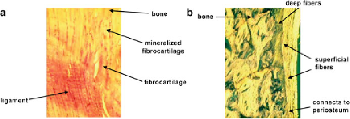
Fig. 4.1
Photomicrograph demonstrating a direct insertion: the femoral insertion of rabbit medial
collateral ligament (
a
), and an indirect insertion: the tibial attachment of the MCL (
b
) (reproduced,
with permission, from [
11
])
4.2.1.1 Direct Insertions
Microscopically, superficial and deep fibers of the ligaments and tendons are inserted
into bone with the deep fibers meeting the bone at right angles with four distinct zones.
Zone 1
consists of type I collagen as the extracellular matrix with fibroblasts. There are
also capillaries, arterioles, and venules running parallel to the collagen fibers, and this
zone is free of nerves and nerve endings. In
Zone 2
, the cells become larger and more
rounded and chondrocyte-like. Cells in this zone are arranged in rows between the
parallel collagen fibers. The Golgi organelles become prominent, and the cell pro-
cesses are short, with most appearing to remain in a lacunar region that extends
1-2
m around the cells. Microscopically, this zone has an appearance similar to
fibrocartilagenous tissues. In
Zone 3
, the tissue becomes mineralized fibrocartilage.
Light microscopy shows a clear line of calcification, called the “tidemark.” The
tidemark is usually smooth in contour, but is sometimes irregularly shaped. About
12
m
m away, the mineral crystals increase in number and aggregate into masses so
that individual crystals become less obvious. The deeper part of zone 3 is also
characterized by dense mineral deposits both within and between the collagen fibers.
Finally, in
Zone 4
, it becomes bone, as the inserting tissuemerges with the bone matrix
collagen fibrils. The bonematrix proteoglycans are represented by chondroitin sulfate,
similar to the small proteoglycans of cartilage, whereas the proteoglycans of ligaments
and tendons are represented by dermatan sulfate.
The femoral attachment of the MCL to the femur is a good example of a direct
insertion. The MCL inserts in the femoral epiphyseal area by passing acutely into
the cortex through a well-defined zone of fibrocartilage and with minimal contribu-
tion into the overlying periosteum (Fig.
4.1a
).
m
4.2.1.2
Indirect Insertions
Unlike direct insertions, the superficial components of an indirect insertion blend
into the periosteum, while the deep components of fibers of indirect insertions
attach to the bone with little or none of the transitional zone of the fibrocartilage
Search WWH ::

Custom Search