Biomedical Engineering Reference
In-Depth Information
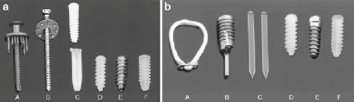
Fig. 13.4 Left
: Tibial side soft tissue devices. A: WasherLoc, B: spiked washer, C: Intrafix, D:
BioScrew, E: SoftSilk, F: SmartScrew.
Right
: Femoral side soft tissue fixation devices. A:
EndoButton, B: Bone Mulch Screw, C: RigidFix, D: Bioscrew, E: RCI Screw, F: SmartScrew
(
Left panel
reproduced, with permission, from ref. [
46
].
Right panel
reproduced, with permission,
from ref. [
47
].)
increased density and number of small diameter collagen fibrils. At 6 months, the
autograft group also had higher ultimate load-to-failure and less anterior tibial
translation [
38
]. In a dog model evaluating fresh soft tissue allograft and autograft
at 6 months, the autograft group demonstrated a more organized four-layer insertion
site compared to a less organized site in the allograft group [
39
].
Most of the studies using animal models to compare allograft vs. autograft use
different processing and sterilization techniques, different graft tissues, and different
methods of evaluating histologic and mechanical properties. This makes drawing
comparisons between studies difficult.
13.4.2 Graft Fixation Techniques
Graft fixation is a critical aspect of ACL reconstruction. Biomechanical testing has
shown that graft materials have higher initial strength than the native ACL [
40
-
42
].
However, multiple studies have shown that by 6 weeks after graft transplantation,
the strength of the graft material is significantly decreased due to the intrinsic
remodeling that takes place within the graft [
43
,
44
]. Therefore, prior to “biologic
fixation” by graft incorporation, the initial mechanical fixation must be secure
enough to allow for early rehabilitation in the first 3 months, which can produce
forces of 450-500 N [
45
].
There are several different fixation techniques that can be used for initial fixation
of soft tissue grafts (Fig.
13.4
). These devices can broadly be divided into suspen-
sory (fixation achieved outside the tunnel) and intra-tunnel (fixation achieved
within the bone tunnel). Multiple biomechanical studies have evaluated the initial
strength of these various devices; however, direct comparisons are difficult due to
the wide variation in fixation and testing methods in these studies.
Even less is known about the influence of these various fixation devices on the
biologic incorporation of soft tissue grafts. In a sheep model, Weiler et al. evaluated
Search WWH ::

Custom Search