Biomedical Engineering Reference
In-Depth Information
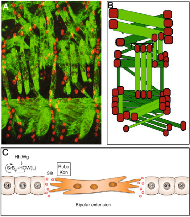
Fig. 6.1
Muscle-tendon interactions in the
Drosophila
embryo. (
a
) Two hemi-abdominal
segments of a stage 16 embryo stained for Myosin Heavy Chain (MHC) (
green
) that marks the
somatic muscles and for Stripe (
red
) marking the tendon cells. Note that each muscle is associated
with two tendon cells at its both ends. (
b
) Schematic representation of a single hemi-abdominal
segment showing the 30 types of muscles (
light green
are anterior muscles and
dark green
are
more posterior muscles, and their tendon attachment cells are in
red
). (
c
) Scheme of the first stage
in tendon assembly; tendon progenitors are defined in the ectoderm by the induction of StripeB
(SrB) by segment polarity genes Hh and Wg. StripeB expression is maintained low as a result of
posttranscriptional repression of the RNA-binding protein HOW(L). SrB regulates positively its
own expression as well as the expression of inhibitor HOW(L). Tendon progenitors secrete Slit
and provide initial cues for directing muscle bipolar migration. The muscle responds to Slit
through Robo receptors. In addition Kontiki (Kon) contributes to the migration of the muscles
(HOW(L)), which is both necessary and sufficient to reduce
stripe
mRNA levels.
HOW(L) itself is a target of StripeB [
18
,
19
]. Thus, HOW(L) creates a negative
feedback loop that counteracts StripeB auto-activation, leading to the maintenance
of StripeB at low levels in the progenitor tendon cells and inhibiting their
subsequent differentiation.
Search WWH ::

Custom Search