Biomedical Engineering Reference
In-Depth Information
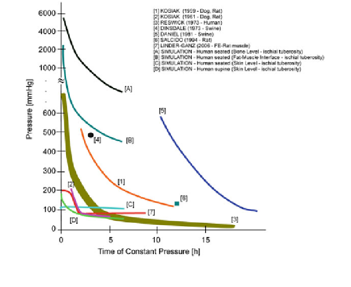
Fig. 6.52 Comparison of simulated tissue direct stress involving an 80 mm soft foam support
with experimental data from other investigations
node-to-node modeling at the fat-muscle and at the muscle-bone interface, tissue
homogeneity, body mass distribution) strongly contribute to tissue stress results.
Simulated tissue stress at the human fat-muscle interface or at the bone level
(Fig.
6.52
/[A] and [B]), however, exceeds that obtained by experimental investi-
gation by more than one order of magnitude! Thus, the use of the above experimental
curves as reference values for pressure sore prevention involving human subjects
becomes questionable (the simulated results indicate that experiments at skin level
are not sufficient, since internal tissue stress apparently drastically increases).
It remains unclear from the above experimental investigations whether cell
death is primarily caused by compressive stress or by shear stress. In most cases,
the location of necrotic tissue, underneath the indenter or lateral to the indenter
edges, where excessive shear deformation occurs, was not specified.
Conclusion: If the empirical pressure-time cell damage relations derived in
animal and human, shown in Fig.
6.52
(Fig.
6.52
/[3] or [7]), are used as a criterion
to evaluate body support systems, the simulated skin level tissue stress values
(Fig.
6.52
/[C] and [D]) are admissible (even if [C] is simulated with respect to a
3 h time span). The simulated stress values at the fat-muscle interface (Fig.
6.52
/
[A] and [B]), however, are not!