Biomedical Engineering Reference
In-Depth Information
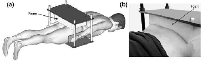
Fig. 6.6 a Test holder with foam specimen attached to top plate and test person in face-down
position--schematic. b Buttock loaded with foam specimen
gel roughly covered the foam surface but did not deeply penetrate the foam cells to
avoid influencing its mechanical properties.
Tablets (Laxoberal
, Boehringer Ingelheim Pharma GmbH, Ingelheim,
Germany) with a high fraction of sodium hydrate, conferring a clearly detectable
signal in the MR-environment served as position markers. They were incorporated
into the foam fixation plate to identify identical positions of FE-model and
MR-images at the stage of comparison.
MRI-scanning was conducted using a conventional 1.5 T system (Magnetom
Espree
, Siemens, Erlangen, Germany). Transversal images of the entire buttocks
(c.f. Figs.
6.9
and
6.10
) of the initially unloaded as well as the loaded configu-
ration were made. Twenty minutes passed after initial tissue loading to allow all
interacting materials to reach steady state before scanning the loaded configura-
tion. This time elapse was based on the findings regarding creep testing with the
specific foam sample material. In addition, conducting creep and relaxation testing
on human gluteal soft tissue showed nearly steady state values at times [5 min.
MRI-settings were chosen as follows: slice thickness -2 mm without gap, field
of view 400 9 450 mm, matrix 456 9 512, a combination of a spine matrix coil
and a Flex multi-channel coil was employed, a T1-weighted spine echo sequence
using transversally oriented integrated parallel imaging was used, repetition/echo
time 550/13 ms.
6.2.3.2 FE-Model Generation
To provide three dimensional surface data of the buttocks at the initial unloaded
state, corresponding MR-images were reconstructed using the image processing tool
Mimics
. A FE-model including bone structure, gluteal muscle groups (assumed as
combined) was built (Fig.
6.7
a-c) upon the reconstructed image data utilizing the
HyperMesh
pre-processor. Both tissue components, fat and muscle, were modelled
using second-order tetrahedral continuum elements. The constitutive Ogden model
for non-linear, hyperelastic, isotropic, slightly compressible materials (3.272)
(cf.
Sect. 3.2.6.4
)
was used to model soft tissue behaviour. First-order hexahedral