Biomedical Engineering Reference
In-Depth Information
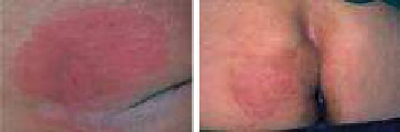
Table 6.1 Classification of pressure ulcers: citations from NPUAP; most wound pictures
adopted from the EPUAP pressure ulcer classification self assessment (Epuap-Puclas 2009)
Grade-I. Intact skin with non-
blanchable redness of a localized
area usually over a bony
prominence. Darkly pigmented
skin may not have visible
blanching; its colour may differ
from the surrounding area
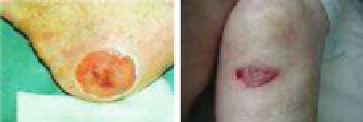
Grade-II. Partial thickness loss of
dermis presenting as a shallow
open ulcer with a red pink wound
bed, without slough. May also
present as an intact or open/
ruptured serum-filled blister
Grade-III. Full thickness tissue
loss. Subcutaneous fat may be
visible but bone, tendon or
muscle are not exposed. Slough
may be present but does not
obscure the depth of tissue loss.
May include undermining and
tunneling
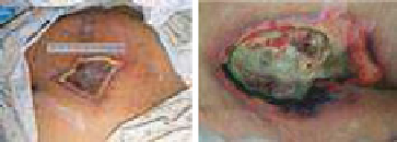
Grade-IV. Full thickness tissue loss
with exposed bone, tendon or
muscle. Slough or eschar may be
present on some parts of the
wound bed. Often include
undermining and tunneling
Suspected Deep Tissue Injury.
Purple or maroon localized area
of discoloured intact skin or
blood-filled blister due to
damage of underlying soft tissue
from pressure and/or shear. The
area may be preceded by tissue
that is painful, mushy, boggy,
warmer or cooler as compared to
adjacent tissue
Unstageable. Full thickness tissue
loss in which the base of the
ulcer is covered by slough
(yellow, tan, gray, green or
brown) and/or eschar (tan, brown
or black) in the wound bed
deep tissue loading due to a body supporting device is necessary to judge effec-
tiveness of the support materials and/or design. By means of simulation methods,
this information can be quantified, visualized and advantageously used towards
design optimization. In addition, to improve interpretation of in vitro and in vivo