Biomedical Engineering Reference
In-Depth Information
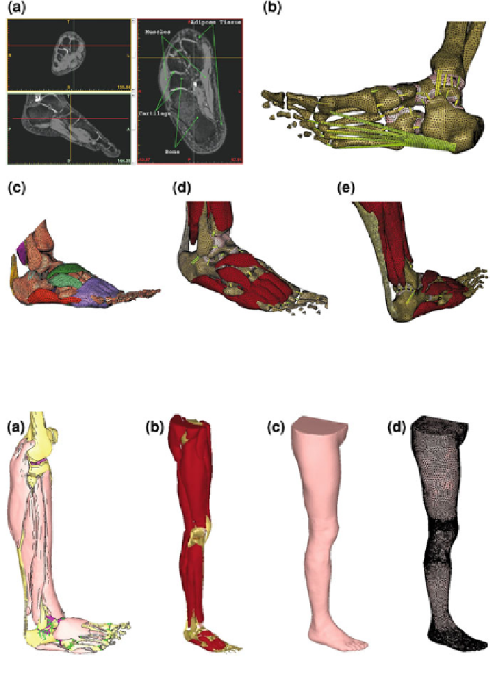
Fig. 5.64 Human foot: a transversal and sagittal MR-scan, b FE-model: bones and major
ligaments, c and d FE-model including bone, muscle and ligaments and A
CHILLES
tendon, e FE-
model: lower leg and foot
Fig. 5.65 FE-model of the human leg: a lower leg including bones, muscles and ligaments,
b complete leg with muscle groups, c skin surface, and d FE-mesh
referred to as a 'gait cycle', whereby each gait cycle is divided into two phases:
(1) stance phase (interval in which the foot is on the ground) and (2) swing phase
(interval in which the foot loses contact with the ground), cf. Fig.
5.66
.