Biomedical Engineering Reference
In-Depth Information
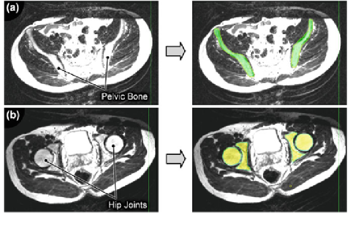
Fig. 5.35 MR image reconstruction based on transversal MRI scans of the human gluteus:
a pelvic bone, and b hip joints
computer tomography (CT) (see Fig.
5.34
a-c). Upright-MRI scanners are avail-
able to scan the human body in the seated or upright position, Fig.
5.34
b.
Prior to scanning, appropriate scan settings, such as sequence or weighting (see
details necessary for image reconstruction. Scanning provides image slices of the
scanned body region. The slice thickness depends on the complexity of the par-
ticular region and the required level of detail. Three orthogonal planes of the
human body are distinguished: sagittal-, transversal- and frontal (cf. Fig.
5.33
):
Sagittal (lateral) plane:
vertical alignment to the sutura sagittalis of the
human skull
Transversal (axial) plane:
horizontal alignment related to the upright body
position and orthogonal alignment related to the
body's longitudinal axis
Frontal (coronal) plane:
parallel alignment to the satura coronalis of the
human skull
In Fig.
5.33
a the planes are shown in relation to the human body, and
Fig.
5.33
b shows the sagittal and frontal plane related to the human skull.
To generate the surface at skin level, it can be appropriate to additionally employ
three-dimensional full body scanning, Fig.
5.34
c.
To obtain a 3D surface representation of the anatomical body regions based on
two dimensional MR scan images, the greyscale-images must be reconstructed