Biomedical Engineering Reference
In-Depth Information
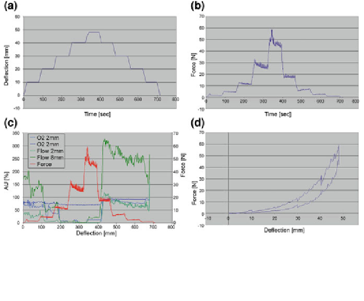
Fig. 5.15 Procedure to evaluate the mechanical properties of gluteal tissue: a displacement-
time-diagram (displacement-driven indenter movement), b force-time-diagram with stepwise
relaxation response, and c in combination with oxygen saturation (blue, light blue) and blood flow
(green, light green) in tissue depth of 2 and 8 mm, and d force-displacement diagram with
termination points
A constant indenter velocity (2 mm/s) was maintained with a total tissue inden-
tation of 44 mm, while indenter force and indentation displacement were recorded,
Fig.
5.15
a. To eliminate frictional effects, the indenter was lubricated. After each
deformation step, a holding time of 2 min was maintained where the indenter
displacement was held constant and the tissue responded with a relaxation process,
cf. Fig.
5.15
b. The force-displacement values reached after each holding period
are referred to as termination points. The force values of the termination points
almost reached a state of equilibrium, cf. Fig.
5.15
b and d. Corresponding ter-
mination points of the loading and unloading paths, cf. Fig.
5.15
d, indicate the
range comprising the pure elastic tissue behaviour (after relaxation) and serve as
basic input for identification of long-term parameters. At maximum indenter
displacement tissue unloading was performed, corresponding to the loading pro-
cedure. The displacement increment sizes (0-5-5-10-10-10-4 mm) of the loading
and unloading cycle were chosen to accommodate the total experimental time,
including seven MR-sessions and test apparatus management. The first two
increments were intended to attain higher resolution of the initial curve slope,
providing minor force increase, whereas the magnitude of the last increment was
due to limitation of the load impact on the test person. The maximum indenter