Biomedical Engineering Reference
In-Depth Information
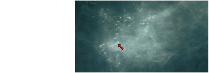
Fig. 13 Mammogram
of a DCIS patient with
characteristic casting-type
microcalcifications,
labeled here with red arrows.
Image courtesy of Andy
Evans, University of Dundee/
NHS Tayside
Models that consider the full spread of time scales in necrosis and calcification can
produce a rich spectrum of behaviors that match observations in pathology [
55
-
57
]
(
Sect. 4
). As hypothesized in [
57
] and investigated in [
56
], fast cell swelling and
lysis—so fundamentally characteristic of early necrosis—are responsible for the
tears (''artifacts'') at the perinecrotic boundary that we consistently see in pathology.
From a continuum point of view, these are rapid perturbations that create persistent
and sharp discontinuities in the cell and necrotic debris distributions.
The simulated tumor microstructure—a viable rim (with greatest proliferation at
the outermost edge) surrounding a stratified, age-structured necrotic core—arises
from the multiscalarity of tissue necrosis and calcification. In the necrotic core, the
structure mirrors tissue age due to the steady flux from the viable rim into the necrotic
core: the newest, least degraded material surrounds increasingly degraded debris,
with central calcifications in the oldest tissues [
56
]. All these features are consistently
observed in patient pathology. Our work revealed a long-time deterioration of cal-
cifications that may explain key features in mammography.
5.1 Next-Generation Hybrid Multiscale Modeling
Improved multiscale and hybrid mathematics and computational techniques are
necessary for further advances. In the agent-based model, each necrotic cell agent
must remain in memory on the order of simulated months; by later times, necrotic
agents outnumber viable agents by three to one or more. And yet the vast majority
of these objects are engaged in the slow time scale processes of calcification and
solid degradation—processes that are well-suited to continuum modeling!
Lowengrub and colleagues are now developing a sophisticated continuum model
of necrotic cell calcification in DCIS [
17
]. We apply a phase field approach [
91
]to
model the tumor as a mixture of fluid, extracellular matrix, and cells. The model can
separately track the necrotic and calcified cell fractions. We also include a sophis-
ticated model of the basement membrane, which can deform in response to
mechanical stresses introduced by the growing tumor [
16
]. Preliminary results
recapitulate the gross features observed in DCIS pathology: a viable rim of appro-
priate thickness surrounding a necrotic core with a calcified center [
17
]. See Fig.
14
.


Search WWH ::

Custom Search