Biomedical Engineering Reference
In-Depth Information
(a)
(b)
(c)
(d)
(e)
Cell volumes throughout
apoptosis
calcification
cytoplasmic solids
cytoplasmic fluids
nuclear fluids
nuclear solids
Cell and nuclear diamete
r vs. time
cell diameter
nuclear diameter
100
20
90
18
80
16
70
14
60
12
50
10
40
8
30
6
20
4
10
0
2
0
2
4
6
8
10
0
2
4
6
8
10
time (hours)
time (hours)
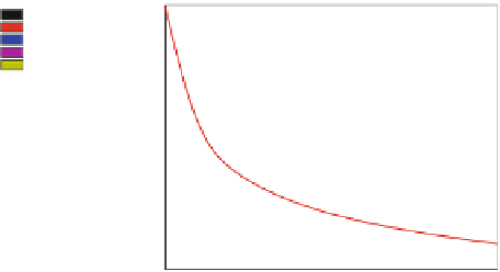
Fig. 2 Apoptosis schematic. Top: a, b While pro-apoptotic signals work to activate initiator
caspases and then effector caspases to degrade subcellular structures and DNA, the cell rapidly
shrinks by removing fluid. c The cell sheds its cytoplasm as membrane-encapsulated blebs.
d, e Chromatin is condensed. DNA is fragmented, encapsulated into apoptotic bodies, and
phagocytosed by nearby cells. Bottom: Preliminary simulation [
64
] of apoptotic cell volume
composition (left) and nuclear/total diameters (right). Figures provided courtesy of [
64
]
mitochondrial membranes are permeabilized and release cytochrome c and other
proteins into the cytoplasm to activate the initiator caspases. In the second, pro-
apoptotic signals directly activate the initiator caspases independently of the
mitochondria [
38
]. Mitochondria-regulated apoptosis disrupts ATP (energy) pro-
duction by decreasing the mitochondrial membrane potential. The cell's remaining
ATP store is depleted by energy-intensive processes throughout apoptosis [
80
]. See
[
65
,
80
] for greater detail on early regulation of apoptosis. While we do not describe
them here, there are also caspase-independent apoptosis mechanisms [
25
,
38
].
After apoptosis is initiated, various ion pumps on the cell's surface quickly
remove water from the cell, resulting in significant volume loss [
6
,
14
,
65
,
67
]. See
Fig.
2
(top: a, b) and Fig.
2
(bottom). Indeed, cell shrinkage and separation from
neighboring cells are some of the first visible signs of apoptosis in histopathology.
The initiator caspases cleave and activate effector caspases (e.g., Caspase-3),
which degrade cellular proteins [
25
,
38
]. The cytoplasm collects in bulbous
''blebs'' that are shed from the cell. See Fig.
2
(top: c). These blebs surround cell
protein
fragments
with
intact
membrane,
and
thus
typically
do
not
trigger
inflammation [
25
,
44
,
65
].
In the nucleus, the chromatin condenses and is henceforth degraded by endo-
geneous endonucleases into short fragments of DNA [Fig.
2
(top: d)]. Protein
cross-linking (e.g., by transglutaminase [
32
]) helps to bundle these fragments into


Search WWH ::

Custom Search