Biomedical Engineering Reference
In-Depth Information
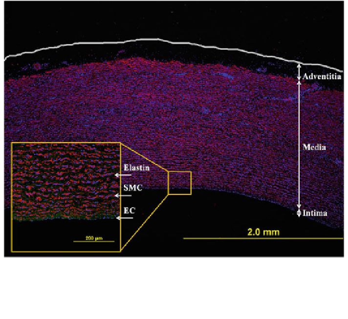
Fig. 1 Porcine aorta displaying the three arterial layers: thin intima, media, and thin adventitia.
The monolayer of endothelial cells is revealed by the blue lining (cell nuclei) in the large image.
The smooth muscle cells (SMC) are shown in red (alpha smooth muscle actin staining) and the
layered elastin in green, which separates each SMC layer. One vessel of the vasa vasorum is seen
in the upper left of the large image as a round collection of SMCs
such as atherosclerosis. Realization that many functions of the endothelium cor-
relate with changes in hemodynamic loads provided important guidance for
treating many vascular disorders and renewed interest in the biomechanics even
though this layer contributes little to the overall structural integrity of the wall.
Flow induced wall shear stresses tend to be on the order of 1.5 Pa in arteries and
0.15 Pa in veins; the mean value of this stress can be estimated in vivo from
measurements of viscosity, volumetric flow-rate, and luminal radius.
The media, or middle layer, consists primarily of SMCs embedded in ECM
consisting of elastic fibers, various types of collagen (I, III, V, etc.), and proteo-
glycans (Fig.
1
). In general, the closer these vessels are to the heart the more
elastin (the main constituent of elastic fibers), and the farther away the more
smooth muscle. Regardless, the mean circumferential wall stress tends to be on the
order of 100 kPa, which can be estimated in vivo via measurements of transmural
pressure, inner radius, and wall thickness. Whereas SMCs primarily synthesize
proteins of the ECM during development and disease, they endow the normal
mature wall with an ability to constrict or dilate and thereby regulate blood flow
locally. Smooth muscle contraction, hypertrophy (increase in size), hyperplasia
Search WWH ::

Custom Search