Biomedical Engineering Reference
In-Depth Information
Unit Cell
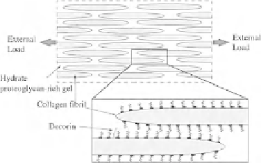
Fig. 8 Collagen fibril micromechanical model [
84
]. A model put fourth for collagen fibers
consisted of fibrils of a finite length and with tapered tips connected via proteoglycan matrix
material (left). A quarter symmetric micromechanical model was defined (right) and subjected to
simulated loading. Figure adapted from [
84
]
using experimental or analytical approaches. Within these studies, the concept of
an inter-fiber matrix material was utilized. The matrix material is thought to
consist of proteoglycans, elastin and other ECM proteins that may mechanically
couple collagen. Such a concept has been used in numerous studies (e.g. [
17
,
18
,
132
,
187
,
232
]) and is used to describe the substance that mechanically couples
collagen fibrils and fibers within tendon and ligament.
One area that shows great promise in the field of multiscale modeling is in the
study of stress and strain localization as it pertains to damage initiation. Although
no studies have yet utilized micromechanical models to study damage initiation in
tendon, they have been utilized in studying microscale strain patterns in the
myotendinous junction (MTJ), which displays similarities to tendon and ligament
tissue. In one such study, a 2D micromechanical model was used to explore
microscale strain distributions within the MTJ, a common location for musculo-
skeletal injuries [
205
]. At the MTJ, muscle fibers taper as they insert into the
tendon via the endomysium, creating a potential location for strain concentration
and damage initiation. By utilizing a microscale unit cell model, this study sought
to investigate strain concentrations within this region. The unit cell consisted of a
single tapered muscle fiber inserting into tendon at a pennation angle of 37
(Fig.
9
). The endomysium was given a transversely isotropic constitutive model
similar to that presented in
Sect. 4.2
and the muscle fiber was given an active
contraction material model developed for muscle tissue [
205
]. The unit cell was
subjected to prescribed displacement along the fiber direction corresponding to a
24 % strain. The edges of the unit cell were given periodic boundary conditions,
which simulated a fiber embedded in macroscopic tissue. Simulations were run
with both passive and active fiber recruitment. Model validation was performed by
comparing the predicted fiber strains to those experimentally measured for relaxed
and strained muscle fibers. More specifically, the deflection of the A-bands within
the muscle fibers were experimentally measured and compared to those obtained
from the FE models (refer to [
205
] for more detail regarding validation methods).


Search WWH ::

Custom Search