Biomedical Engineering Reference
In-Depth Information
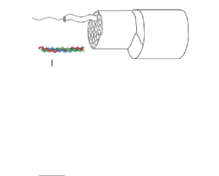
Ligament
Fascicle
Fiber
Fibril
Tropocollagen
Nano
100 nm
Micro
100 µm
Meso
500 µm
Macro
5mm
Fig. 1
Hierarchical organization of ligament from the molecular level to the joint level
fibers form fascicles at the mesoscale and fascicles form the whole tendon or
ligament at the macroscale (Fig.
1
).
The ECM of ligaments and tendons is formed by self-assembly of cell-secreted
proteins and consists of approximately 70 % water [
28
]. The solid phase of these
tissues is primarily composed of type I collagen (60-80 %), with the remainder
consisting of elastin, proteoglycans and glycosaminoglycans (GAGs), other types
of collagen (types III, IV, V, VI), fibrillin and other proteins [
121
,
125
,
230
]. Type
I collagen exhibits different organizational motifs at each scale (Figs.
1
and
2
)
[
121
]. At the nanoscale, tropocollagen monomers are assembled to form fibrils
(50-200 nm dia.), which display a characteristic d-banding period (67 nm) [
121
,
170
,
171
,
224
]. Tropocollagen monomers are held together by a combination of
hydrogen, ionic and covalent bonds [
125
,
230
]. Fibrils are spaced regularly within
healthy tissue and predominantly aligned in parallel [
31
,
209
,
227
]. Cross sectional
TEM images reveal that fibrils are well organized and separated by a regular
spacing within healthy tissue. At the
microscale
, fibrils are assembled into fibers
(20-50 lm dia.) [
50
,
117
]. Fibroblasts and tenocytes (10 lm width 9 60 lm
length) are located in the interfiber space [
121
,
125
]. Fibroblasts and tenocytes are
responsible for regulating the ECM in response to loading and injury, and
mechanotransduction plays a major role in their function [
229
]. The characteristic
crimp pattern is visible at the fiber level, with a period of 50-200 lm[
112
,
117
].
Fibers are arranged in a largely parallel fashion [
121
]. At the
mesoscale
, fibers are
assembled into fascicles (100-500 lm dia) [
50
,
121
,
230
]. To at least some extent,
crimp is registered between fibers [
124
,
168
]. Fascicles are organized in parallel
[
96
]. Fascicles and fibers are surrounded by a thin fascia (referred to as endotenon)
[
85
,
121
,
206
]. At the
macroscale
, groups of fascicles are organized into functional
bands (100 lm-1 mm dia) [
50
].
Noncollagenous ECM constituents include proteoglycans (PGs) such as decorin
(*1%/wt), biglycan (*0.5%/wt) and others (fibromodulin, lumican, aggrecan,
versican), fibrillin [
21
,
114
,
125
,
143
-
145
,
167
] and elastin (1-2%/wt) [
121
,
125
,
192
,
206
]. The large PGs (e.g., aggrecan) contribute to the apparent viscoelastic
material behavior of these tissues by controlling water content and flux [
114
].


Search WWH ::

Custom Search