Biomedical Engineering Reference
In-Depth Information
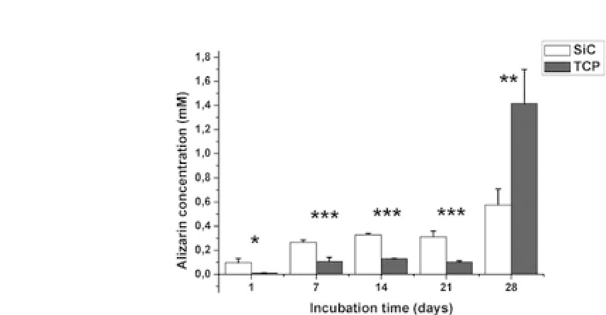
Figure 2.7.
Quantification of calcium deposits, stained by Alizarin Red
over the bioceramics and TCP, up to 28 days of cell culture. (Statistical sig-
nificant differences:
∗
p
<
0
.
05
,
∗∗
p
<
0
.
01, and
∗∗∗
p
<
0
.
005)
.
Once the osteoblastic cells covered the whole surface and pores
ofthesapelli-basedceramics,theystartedtosynthesizetheextracel-
lularmatrix,followedbythedepositionofcalciumphosphate.These
calciumdepositsweredeterminedbyAlizarinRedstainingandsub-
sequentquantification(Fig.2.7).Calciumdepositsrevealedahigher
and earlier level of mineralization on SiC ceramics than on TCP dur-
ing the first 21 days of incubation. Calcium deposits were detected
inside the channels of the SiC ceramics even at 7 days of incuba-
tion and over and around the ceramics at 14, 21, and 28 days. These
results confirmed that the surface microstructure of the SiC favored
the earlier differentiation of the osteoblasticcells.
Inadditiontothisassay,andtoconfirmthepresenceoftheextra-
cellular matrix and the mineralization process, SEM was used to
evaluate the ceramics at 28 days of incubation (Fig. 2.8). The SEM
micrographs show the whole surface of SiC covered by a thick layer
of cells (Fig. 2.8a). When the surface was analyzed closely, a pre-
mineralized extracellular matrix was observed over the cell layer
(Fig. 2.8b-e). Collagen fibers, oriented in all directions to form the
trabecular bone,were also found(Fig. 2.8f).
It has been previously reported
23
,
24
that the surface microstruc-
ture of different materials has a great influence on the cellu-
lar acceptance of the materials. Structural features of the surface


Search WWH ::

Custom Search