Biomedical Engineering Reference
In-Depth Information
theseconditions,FGF-2aloneisabletostimulatesubstantialgrowth
of MSCs. However, the combination of FGF-2 and dexamethasone
induces a much higher increase in MSC proliferation. As is also
observed in 2D culture, dexamethasone alone arrests MSC growth
in the 3D scaffold. These results suggest that, at least in the case
of FGF-2 and dexamethasone, growth stimulation of MSCs can be
reproduced inboth 2D culture and 3D scaffold culture conditions.
Stimulation of MSC-loaded BCP scaffolds with FGF-2 and dex-
amethasone for 7 days, followed by stimulation with osteogenic
medium (50 mg/mL ascorbic acid, 10
-
2
M
β
-glycerophosphate, and
10
-
8
M dexamethasone) for an additional 14 days, significantly
inducedinitiationofdifferentiationtowardtheosteoblastlineageas
assessedbyALPstaining(Fig.41.2).Theseresultsdemonstratethat
osteoblast commitment is also increased in MSCs grown in a scaf-
foldwithFGF-2anddexamethasone.Theenhancementofosteogenic
differentiation by FGF-2 and dexamethasone may be in part due to
the high proliferation rate of MSCs in 3D culture. Because inhibitors
of Src kinase suppress both proliferation and osteogenic differen-
tiation, and the degree of osteogenic differentiation in 2D culture
is dependent on cell number,
43
the enhanced proliferation rate of
MSCsin3DmaycontributetotheosteogenicdifferentiationofMSCs
expanded with FGF-2 and dexamethasone. It is also possible that
osteoblast commitment may contribute to enhanced osteogenic
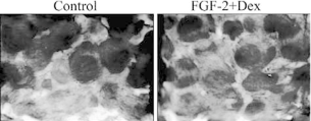
Figure 41.2.
ALP staining of MSCs treated with FGF-2 and Dex in a BCP
scaffold. Bone marrow MSCs (2
×
10
5
cells) were seeded onto a porous
BCP ceramic scaffold, cultured with or without a combination of FGF-2 (1
ng/mL) and Dex (10
−
8
M) for 7 days, and then treated with an osteogenic
cocktail for an additional 14 days. Cells committed to the osteoblast lineage
were identified by ALP staining.


Search WWH ::

Custom Search