Biomedical Engineering Reference
In-Depth Information
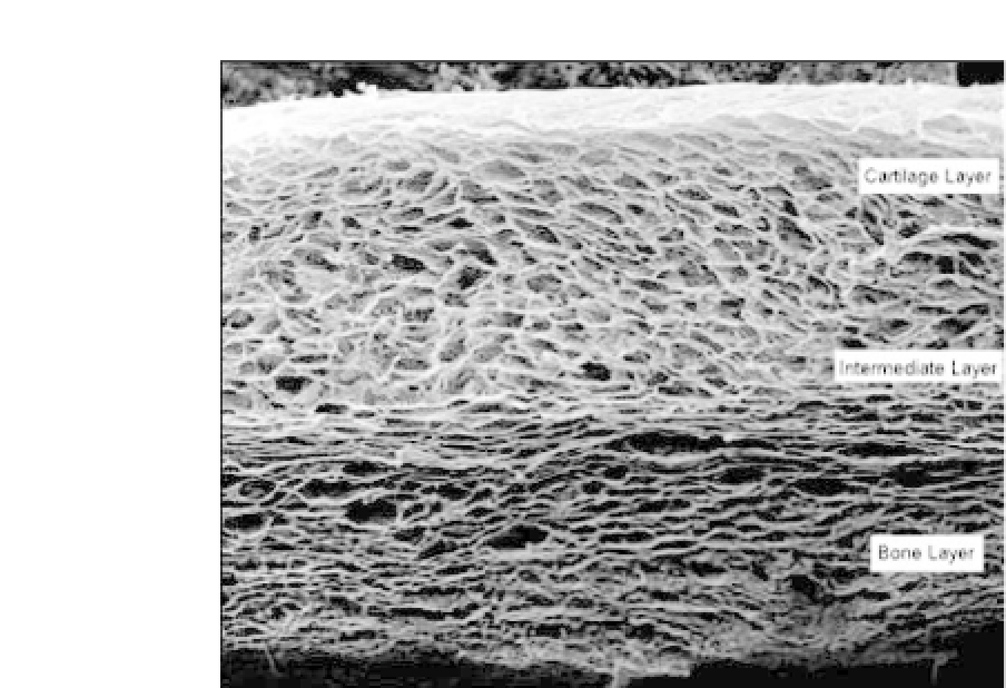
Figure 38.2.
SEM image of ChondroColl, which is a 3-layered osteochon-
dral scaffold composed of a bone layer (containing type 1 collagen and HA),
an intermediate layer (containing type 1 collagen, type 2 collagen, and HA),
and a cartilage layer (containing primarily type 2 collagen).
Abbreviation
:
SEM, scanning electron microscopy.
throughout the structure to promote tissue formation by different
cell types throughout thestructure.
Previously we have shown
61
that scaffold pore size significantly
affects cellular activity, and it is hypothesized that the optimal scaf-
fold pore size is not only tissue specific but also cell specific. For
example, it is hypothesized that scaffolds for bone tissue engineer-
ing should contain pores
>
300
μ
m to enhance bone formation
and vascularization,
62
while scaffolds for cartilage tissue engineer-
ing should employ a pore size
<
200
μ
m.
63
-
65
It is thought that
smaller pores sizes may encourage hypoxic conditions, which are
more favorable for chondrogenesis. Therefore, by producing a scaf-
fold with a different mean pore size in the osteo and chondral
regionsorbyproducing ascaffoldwithagradientinmeanpore size
fromtheosteotothechondralregion,itmightbepossibletodevelop
an optimal scaffold for osteochondral repair.


Search WWH ::

Custom Search