Biomedical Engineering Reference
In-Depth Information
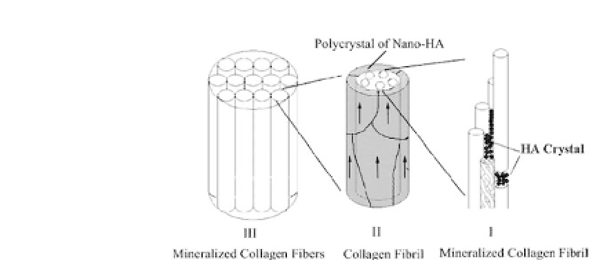
Figure 37.4.
Schematic diagram of the hierarchical structure of self-
assembled HA-collagen composite.
44
(I) The first level of the hierarchy is
the organization of collagen molecules with the nano-HA crystals formed
initially in the gap zones between the collagen fibrils. (II) The second level
of the hierarchy is the organization of collagen fibrils with respect to HA
crystals. The HA crystals are sheetlike and grow on the surface of these fib-
rils in such a way that their c-axes are oriented along the longitudinal axes
of the fibrils, as indicated by the arrows in the figure. (III) The third level
of the hierarchy is the organization of the mineralized collagen fibrils. The
mineralized collagen fibrils align parallel to each other to form mineralized
collagen fibers.
A schematic diagram of self-assembled HA/collagen composites
comprising multiple levels of hierarchical organization has been
depicted in this study. As shown in Fig. 37.4, the lowest level of this
hierarchy is the organization of collagen molecules with some par-
ticulatesofnano-HAcrystals.Thecollagenfibrilsareformedbyself-
assembly of collagen triple helices, and the HA crystals are formed
initiallyinthegapzonesbetweenthecollagens.Consideringthatthe
diameter of the collagen molecule is 1.5 nm, the diameter of the col-
lagen fibrils in the five-stranded packing model should be approx-
imately 4.0 nm, which is in agreement with TEM observations of
self-assembled collagen fibrils. The second level of the hierarchy is
the organization of these collagen fibrils with respect to the growth
of the HA crystals. HA crystals grow on the surface of these fibrils
in such a way that their c-axes are oriented along the longitudinal
axesofthefibrilsandsurroundthefibrils.Thisarrangementimplies
that the nucleation and growth of HA crystals are not random but
rather are controlled by the fibrils themselves. The third level of the


Search WWH ::

Custom Search