Biomedical Engineering Reference
In-Depth Information
Figure 35.1.
Phasecontrastphotomicrographshowingthemorphologyof
the cultured gingival fibroblasts.
8-12 T-225 flasks (7-10 days). Cells cultured beyond the sixth pas-
sage were notused for treatments.
The characteristics of the cultured cells were checked by
immunofluorescence microscopy for known fibroblast markers
(Fig.35.2).Thecellswereseededina6-wellplate(GreinerBio-One)
at a density of 2,500 cells/cm
2
and cultured until the cells became
subconfluent.CellswerewashedthreetimeswithPBS(NissuiPhar-
maceutical Co.) and then fixed with 4% paraformaldehyde (Sigma-
Aldrich) for 30 minutes. Cells were washed three times with PBS
for 5 minutes and treated with a blocking reagent containing 2%
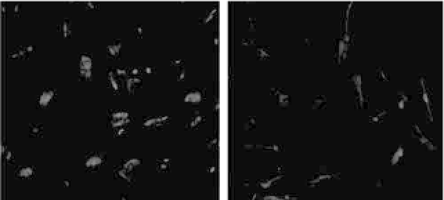
Figure 35.2.
Dark-field photomicrographs showing results of indirect
immunofluorescent staining of cultured gingival fibroblasts. Nuclei (blue)
were visualized by counterstaining with DAPI. (a) Staining with anti-
collagen type I and (b) staining with antivimentin. See also Color Insert.


Search WWH ::

Custom Search