Biomedical Engineering Reference
In-Depth Information
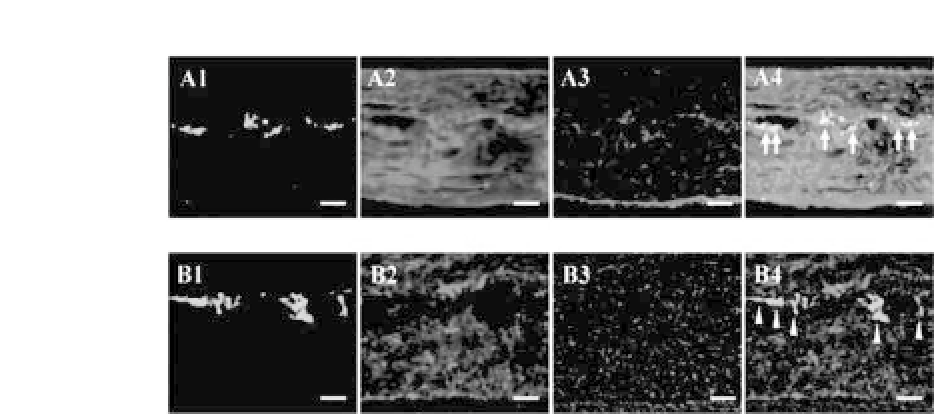
Figure 34.16.
Phenotypic switch of dermal fibroblasts to corneal stromal
cells. After 8 weeks of implantation, keratocan expression is observed in
the group with the implantation of fibroblast-PGA constructs precultured
in vitro
for 1 week (a, arrow) but not in the group with 3-week precultured
cell-PGA constructs (b, arrow head). a1, b1: GFP expression; a2, b2: kerato-
canstaining;a3,b3:nucleistaining(Hoechst33258);a4,b4:mergedimages
of 3 colors. Bars: 50
μ
m. (Reprinted by permission from Ref. 19). See also
Color Insert.
new tissue environment under restricted conditions. The functional
restoration of corneal transparency using dermal fibroblasts sug-
geststhattheycouldbeanalternativecellsourceforcornealstroma
engineering.
19
34.6 PGA Fibers for Blood Vessel Engineering
To prove the possibility that a neovascular structure containing
both endothelium and a smooth muscle (SM) layer can be engi-
neered, studies were performed first in a nude mouse model.
Endothelialcellsandsmoothmusclecells(SMCs)wereisolatedfrom
human neonate umbilical veins. After
in vitro
expansion, these cells
were seeded onto unwoven PGA fibers wrapped around a silicone
tube and then
in vitro-
co-cultured for one week followed by
in
vivo
implantation into the subcutaneous tissue of a nude mouse. A
scaffold tube alone was transplanted as a control. Engineered ves-
sels were harvested at 2, 6, and 11 weeks postrepair for histol-
ogy and immunohistochemical staining. Grossly, a tubular structure


Search WWH ::

Custom Search