Biomedical Engineering Reference
In-Depth Information
material has also been used in skin engineering. Briefly, autologous
full-thickness skin (2
×
2 cm) was harvested, and keratinocytes and
dermal fibroblasts were isolated and then separately seeded onto
a sheet of unwoven PGA fibers with a polyethylene-polypropylene
hydrogel. Two layers of cell-scaffold sheets were then overlapped
to form a composite skin construct with a thickness of 2 mm. In a
porcine model, two full-thickness wounds (4 cm in diameter) deep
to the fascia were created at the dorsal aspect of the pigs, and a
titanium isolation chamber was then inserted to prevent tissue in
growth from adjacent skin. The wounds were repaired either with a
composite skin construct as an experimental group or with bioma-
terial alone as a control group. The engineered skin was harvested
at one, two, four, and eight weeks postrepair for histological exam-
ination. When examined grossly, neoskin formation was observed
at two weeks postrepair in the experimental group. Although the
woundwastotallycoveredbytheengineeredskinintheexperimen-
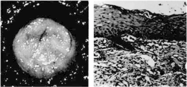
talgroupatfourweeks,mature, full-thicknessskinwasformedonly
in part of the wound area. However, the maturation completed uni-
formly at eight weeks, which resembled the morphology of native
porcine skin (Fig. 34.12,
left
).
In contrast, only granulation tissue was observed in control
woundsat differenttimepoints. Histologically, adouble-layerstruc-
ture was observed as early as one week postrepair. Interestingly,
the rete ridges of the epidermis became enlarged and started to
migratedeeplyintodermalpartoftheengineeredskinattwoweeks
and reached further deeper at four weeks. At eight weeks, these
Figure 34.12.
Gross view (
left
) and histology (
right
) of engineered full-
thickness skin at 8 weeks. (Reprinted by permission from Ref. 8).


Search WWH ::

Custom Search